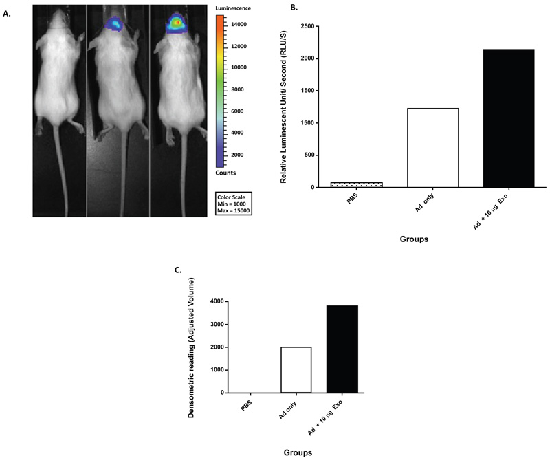
Fig. (2) Mouse NSC-derived exosome-mediated in vivo enhancement of Ad infection (unpublished work by Matthews et al.). (A) Mice were injected with PBS, 1x10⁸ Viral Particles (VP) of Ad, or 1x10⁸ VP of Ad co-incubated with 10 µg of exosomes. At 24 hours, post-injection mice were imaged by means of non-invasive luciferase imaging. The imaging results were represented as total luminescent counts. (B) Quantitation of representative mice from Fig. (2A), data represented as relative luminescent counts per second. (C) Densitometric analysis of western blot analysis of mouse brains at 48 hours post-injection (PBS, 1x10⁸ VP of Ad, or 1x10⁸ VP of Ad co-incubated with 10 µg of exosomes). Anti-adenovirus antibody was used for western blot analysis.