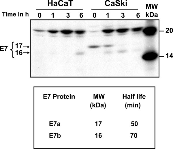
Fig. (2) Processing and half-life of the HPV-16 E7 protein. HaCaT and CaSki cells were pulse labeled with [35S]-methionine-cysteine for 15 min and chased for different times (0, 1, 3 and 6 h). Upper panel: Cell extracts were immunoprecipitated with the anti-E7 polyclonal C89 antibody, samples separated by SDS-PAGE gel and bands visualized by auto-radiography. The arrows showed a 17 kDa (E7a) and a 16 kDa (E7b) bands. The 15 kDa band observed in HaCaT cells is a non-specific band as it is not consistently observed in experiment repetitions. Lower panel: Bands were scanned and plotted in a graph to calculate the half-life of the different forms of E7 protein.