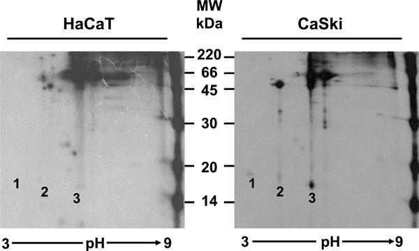
Fig. (3) Characterization of the HPV-16 E7 proteins by IEP. HaCaT and CaSki cells were labeled with [35S]-methionine-cysteine and immunoprecipitated with the C89 polyclonal antibody. The immunoprecipitates were separated in the first dimension in a pH gradient from 3-9 for 16 h. The second dimension was run in a 15% PAGE gel and treated for fluorography. The numbers over the CaSki panel show the localization of the 3 isoforms of HPV-16 E7 proteins (E7a1, E7a and E7b) and these spots were not observed in the HaCaT control cells.