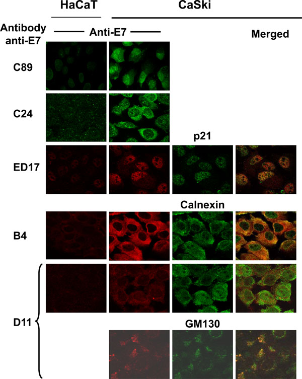
Fig. (5) Subcellular localization of HPV-16 E7 proteins by immunofluorescence. Cells were fixed with p-formaldehyde and permeabilized with 0.2% saponine. Cells were tested with the different anti-E7 polyclonal (C89 and C24) and mAbs (ED17, B4, D11). Biological cell markers were used to identify ER (calnexin), Golgi (GM130) and nucleus (p21) and co-localized with the immunological detected E7 protein. Secondary fluorescent antibodies were anti-rabbit IgG conjugated with Alexa 488 (green) or anti-mouse IgG conjugated with Alexa 594 (red). The merged image is shown in the right side of the figure and positive co-localization is observed in yellow color. Images were taken at a magnification of 1000X using Confocal microscope.