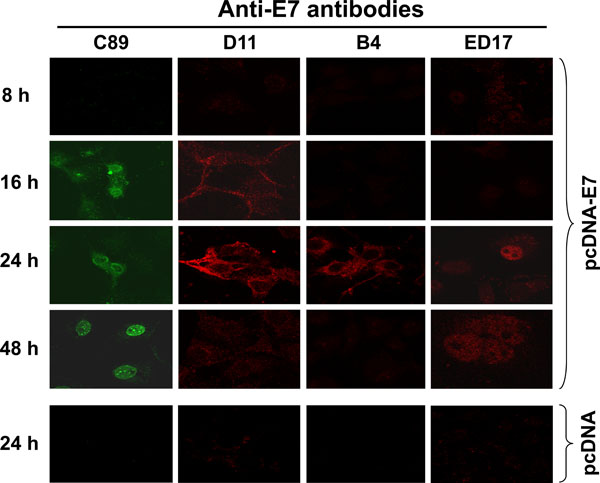
Fig. (6) Processing of the HPV-16 E7 protein under in vivo conditions. Cos-7 cells were transiently transfected with the pcDNA-E7 plasmid and chase for different periods of time (8, 16, 24 and 48 h). Harvested cells were fixed with 4% p-formaldehyde, permeabilized as described in Material and Methodology and tested with different anti-E7 antibodies. The antibodies tested were C89 polyclonal antibody, and D11, B4 and ED17 mAbs. Secondary fluorescent antibodies were anti-rabbit IgG conjugated with Alexa 488 (green) or anti-mouse IgG conjugated with Alexa 594 (red). Cos-7 cells transfected with pcDNA plasmid alone at 24 h was used as control of the system as this was the time for the highest expression of the E7 protein observed. Images were taken at a magnification of 1000X using Confocal microscope.