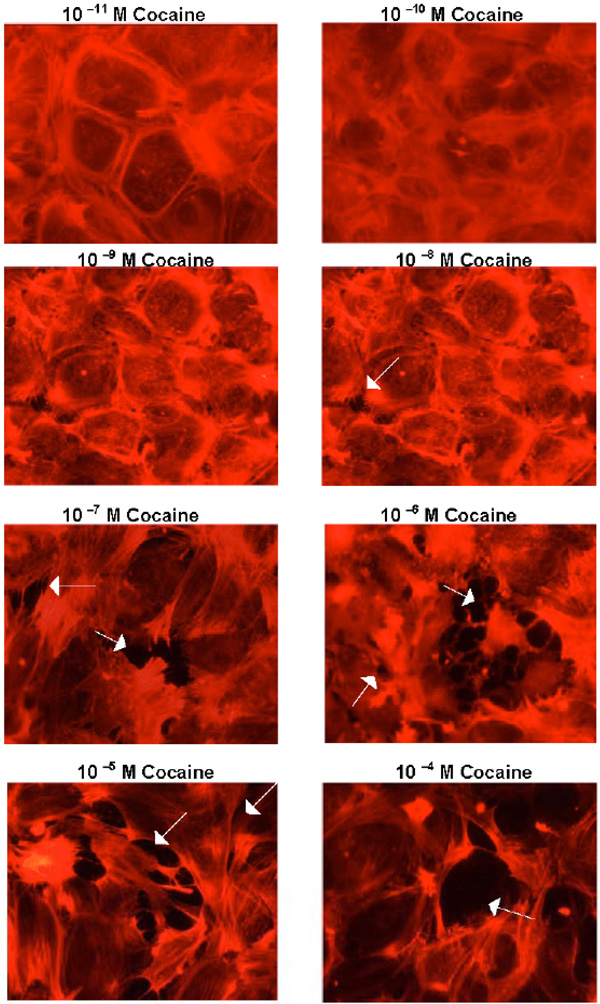
Fig. (1) Cocaine disrupts BMVEC junctions. Confluent BMVEC monolayers were treated with the indicated concentration of cocaine for 1h; the cells were then washed, fixed, stained with phalloidin-ALEXA 594 and photographed by Hamamatsu camera on the Olympus Bmax microscope (40x). The arrows indicate gaps between BMVEC’s.