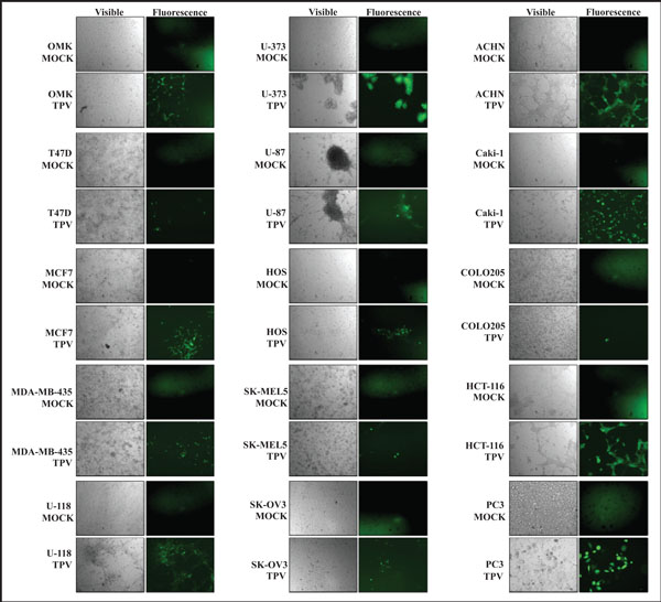
Fig. (1) UV fluorescent microscopy of TPV-GFP infected cancer cells at 7 days post infection. Left panels in each case show cell monolayers under visible light. Right panels show same area under UV fluorescence. Uninfected cells (MOCK) show no green fluorescence while TPV-GFP infected plaques emit green fluorescence. Greater progression of TPV-GFP infection is observed in U-373, HCT-116, ACHN, and Caki-1cell monolayers when compared to the OMK control.