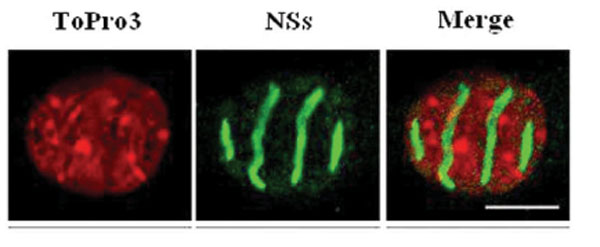
Fig. (2) RVFV NSs forms a filamentous structure in the nucleus. Confocal microscopy picture showing a section of a L929 cell nucleus infected by ZH548 and stained with the DNA intercalating dye ToPro3 (red), with anti-NSs antibodies (green) and merge. The cellular DNA is predominantly excluded from the filament (from Mansuroglu et al., J. Virol. in press).