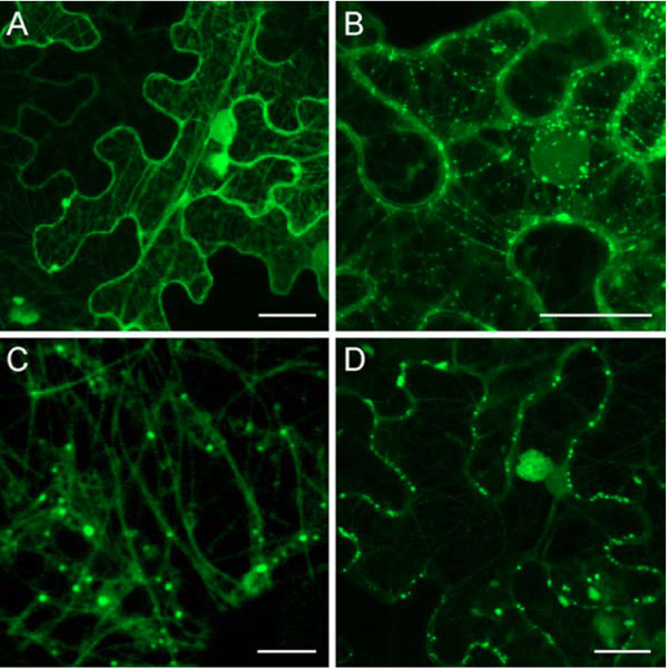
Fig. (3) Localization of PMTV TGBp1 fused to GFP in epidermal cells of N. benthamiana leaves agroinfiltrated with the construct 35S:GFP-TGBp1-NOS:TGBp2/TGBp3. Cells were imaged at 2 days post infiltration (dpi) (A), 3 dpi (B and C) and 4 dpi (D). The images, except C, are reconstructed by superposition of series of confocal optical sections. C represents a single optical section in the cortical cell region. Scale bars: A, B and D, 20 µm; C, 5 µm.