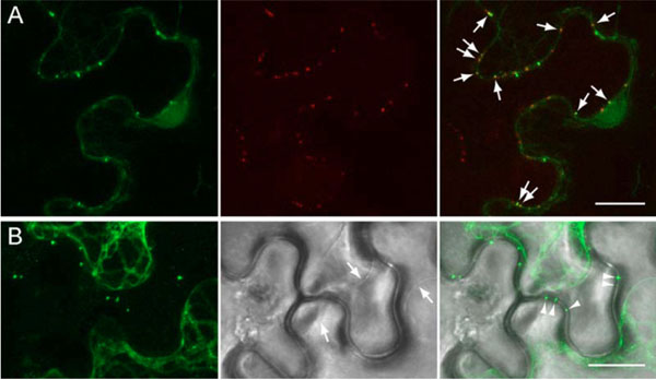
Fig. (4) Co-localization of GFP-TGBp1 with cell wall-embedded plasmodesmata structures in epidermal cells of of N. benthamiana leaves agroinfiltrated with the construct 35S:GFP-TGBp1-NOS:TGBp2/TGBp3. A, GFP-TGBp1 co-localization with callose stained with sirofluorcontaining aniline blue dye; GFP signal is shown in the left panel, callose signal (the middle panel) was digitally pseudocolored with red to facilitate interpretation of the merged image (the right panels). Arrows in the right panel point to overlapping signals. B, GFP-TGBp1 localization in cells subjected to plasmolysis by mannitol treatment. Left panel shows the GFP signal, the middle panel - a bright field image, the right panel - superposition of the images. Arrows show the plasma membranes retracted from cell walls. Arrowheads point to GFP-TGBp1- containing structures retained in the cell wall. Scale bars, 15 µm.