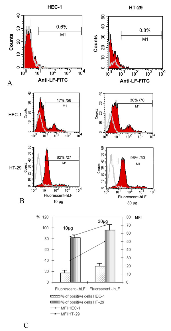
Fig. (1) Binding of hLF to epithelial HEC-1 and HT-29 cells. (A): Lack of constitutive expression of LF at cellular surface of epithelial cells. Cells were incubated with anti-lactoferrin for 30 min at 4°C, washed and incubated with Oregon-fluorescent-conjugated anti-rabbit for additional 30 min at 4°C. Cells were then fixed by PFA (1%) and analysed by FACScalibur analysis. One representative of 4 ruled out independent experiments is shown. (B): Human LF binds to HEC-1 and HT-29. Epithelial cells were incubated with fluorescent hLF at 10 and 30 µg (e.g. 128 and 384 nM) for 1 h at 4°C, fixed and analysed by FACScalibur. The corresponding percentages of positive cells and mean fluorescences are mentioned in the correspond quadrant. Data are from a typical experiment representative of four independent experiments. (C): The mean ± SD of LF positive cells and corresponding mean MFI calculated from 4 experiments is presented.