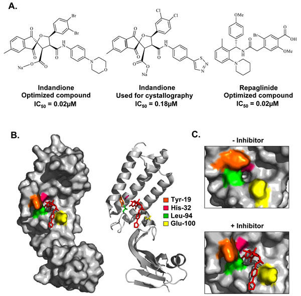
Fig. (4) Inhibition of the E1-E2 protein interaction. (A) Structures and potencies of optimized indandione and repaglinide inhibitors of the E1-E2 protein-protein interaction. (B) Surface and ribbon representation of the HPV11 E2 TAD-indandione inhibitor complex (PDB accession number 1R6K [101]). The structure of the inhibitor used for crystallography is shown in (A). The amino acid residues that form the hydrophobic inhibitor-binding pocket are depicted in stick representation and are colored according to the legend in the figure. (C) Enlarged view of the hydrophobic pocket in the absence (left panel) and presence (right panel) of inhibitor, displaying the significant movement of the amino acids Tyr-19, His-32, Leu-94, and Glu-100 upon inhibitor binding.