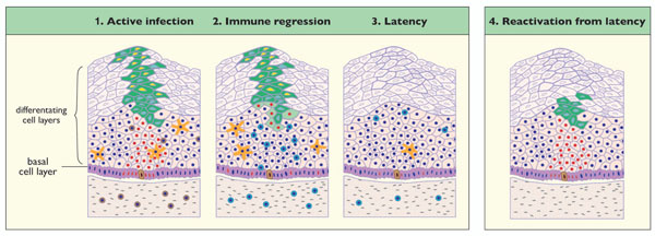
Fig. (2) Model of Papillomavirus Latency based on Animal Studies. (1) Active Infection. Active papillomavirus infection involves the
regulated expression of viral proteins as cells containing viral genomes migrate towards the epithelial surface. The onset of viral genome
amplification (light blue) and L1 expression (yellow) facilitates the assembly and eventual release of virions from the epithelial surface. Cells
that are ‘in the cell cycle’ and which may proceed through cell division are marked with red nuclei. In the upper epithelial layers, cell cycle
entry is driven by the viral E6 and E7 proteins. The virus-infected cells of the epithelial basal layer maintain the viral genome as a low copynumber
episome with only low-levels of viral gene expression. Long-term persistence may require the maintenance of the viral genome in an
epithelial ‘stem cell’ (highlighted in the basal layer). Resting T-cells (brown) and Langerhans cells (orange) can be found in the lower layers
of the epithelium and in the dermis. The positions of these resting T-cells (brown circular cells) are indicated in the diagram. (2) Immune
Regression. Immune regression involves the presentation of viral antigens to the immune system, (probably via the Langerhans cells) and
the subsequent accumulation of activated CD4+ and CD8+ T-cells (light blue circular cells) in and around the lesion. During regression,
activated T-cells accumulate within and beneath the lesion. (3) Latency. Lesion clearance involves the suppression of viral gene expression
as lymphocytes infiltrate, and may involve changes in cytokine activity and cytokine signaling at the site of regression. The ongoing
proliferation of virus-containing basal cells in the absence of normal viral gene expression appears to underlie lesion clearance. Changes in
viral gene expression in the replicating basal cells may explain the slow decline in viral genome copy number over time at the site of
previous infection. Current thinking suggests that the papillomavirus genome may persist long-term in the slow-cycling stem cells or stemlike
cells. (4) Reactivation from Latency. This model may explain the changes in viral copy number that are thought to accompany
immunosuppression. The presence of memory T-cells circulating in the epithelium prevent extensive viral gene expression and keep the viral
genomes in the basal layer in a latent state. Changes in immune status would allow local rises in viral copy-number, and the possible reappearance
of visible papillomas.