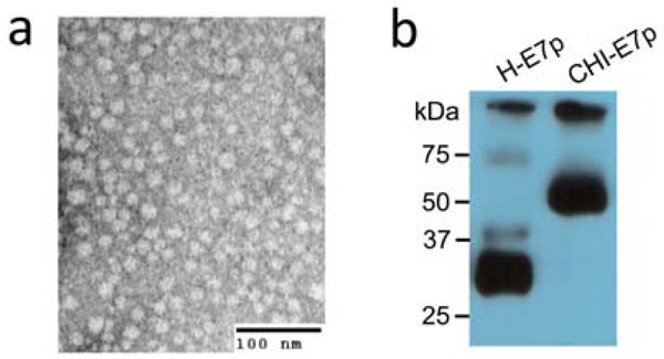
Fig. (2) Analysis of CHI-E7p protein expressed in yeast and
human cells. (a) Transmission electron microscopy of negatively
stained CHI-E7p VLPs purified by ion metal affinity
chromatography (Ni-NTA-agarose columns) from the yeast P.
pastoris transformed with the pPICZ(A)-CHI-E7p plasmid. The
VLPs appear as nanoparticles of 20-25 nm in diameter. (b) Western
blot of HEK293 cells transfected transiently with pcDNA3
plasmids encoding either the CHI-E7p or the H-E7p fusion
proteins. The cells were lysed and electrophoresed 48 hours after
transfection. The proteins were detected with anti-6x His
antibodies. The CHI-E7p protein runs with a molecular weight of
~55 kDa while the H-E7p protein appears as a ~28 kDa band.