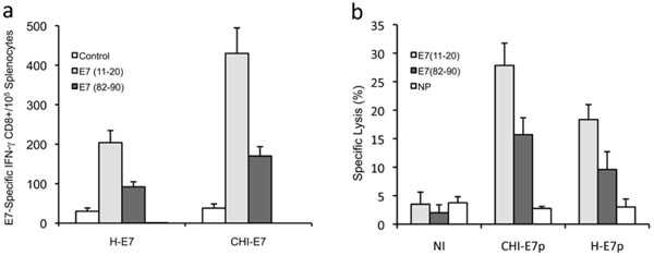
Fig. (4) Detection of HPV16 E7-peptide-specific T cell responses to immunization with plasmid DNA encoding CHI-E7p or H-E7p fusion
proteins. (a) Intracellular cytokine staining and flow cytometry analysis of E7 peptide-specific CD8+ T cell responses. Groups of female
HLA-A2 (AAD) transgenic mice (5-6/group) were immunized with pcDNA3-CHI-E7p or pcDNA3-HE-7p. Splenocytes were isolated and
incubated overnight either with peptide E7(11-20) or E7(82-90) or without peptide (control). After staining with anti-CD8 and anti-IFN-γ
antibodies, the T cells were analyzed by flow cytometry to detect CD8+/IFN-γ+ T cells. The graph shows mean numbers (with s.d.) of IFN-
γ+/E7E7 peptide-specific CD8+ T cells detected per 105 T cells. (b) Cytotoxic T cell responses induced by immunization against the CHI-E7p
or H-E7p fusion proteins as determined by antigen-specific kill of indicator cell populations. Female HLA-A2 (AAD) transgenic mice
were immunized at 2-week intervals with pcDNA3-CHI-E7p or pcDNA3-HE-7p. Six days after the fourth boost immunization, splenocytes
isolated from naïve mice were loaded with peptide E7(11-21) or E7(82-90) or left without peptide (NP), and were subsequently labeled with
CFSE at high, low and intermediate concentrations, respectively. Equal numbers of labeled cells were mixed, and 15 x 106 cells of the
mixture were injected intravenously into control (non-immunized, NI) and immunized mice. Lymphocytes from spleens and regional lymph
nodes were taken 20 h after injection and analyzed by flow cytometry. The data shown are representative of two experiments performed.