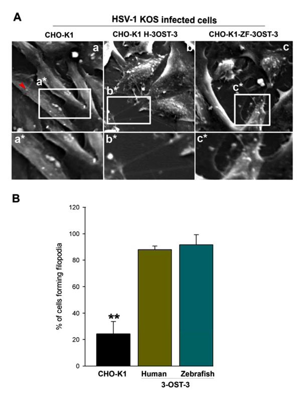
Fig. (1) Role of filopodia during HSV-1 entry into Chinese hamster ovary (CHO-K1) cells expressing Zebrafish (ZF) encoded 3-
OST-3 receptor. (A) Scanning electron microscopy (SEM) performed on HSV-1 (KOS) infected glycoprotein D (gD) receptor negative
(wild-type CHO-K1 cells; panel a) and gD-receptor positive cells (panel b and panel c). CHO-K1 cells expressing human 3-OST-3 (H-3-
OST-3), and zebrafish (ZF) encoded 3-OST-3 (ZF-3-OST-3) were grown at a confluence of 40% in a chamber slides (Lab-Tek chamber
slide) and were exposed to HSV-1 (25 pfu/cell for 45 min at 37°C). The infected cells were fixed with 2% formaldehyde/4% glutaraldehyde
in PBS before SEM. Highlighted regions showing virions bound to wild-type CHO-K1 cells (panel a*) were unable to induce filopodia (red
arrow indicates single Filopodia in panel a), while large number of filopodia (panels b* and c*) were observed in human and ZF encoded 3-
OST-3 expressing CHO-K1 cells. The arrow indicates presence of virus on the induced filopodia. (B) Determination of percentage of
filopodia scored from sampled groups of 100 single cells or clusters (with 5-20 cells) in wild-type CHO-K1 cells, and CHO-K1 cells
expressing human and ZF 3-OST-3 receptor in triplicate experiments, 5 μm length of a protrusion and at least 10% of the cell surface
covered with 25 or more protrusions is scored positive. ** P< 0.05, one way ANOVA.