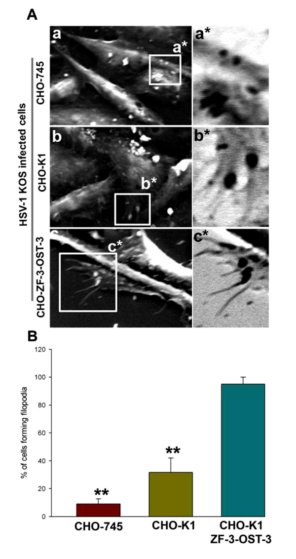
Fig. (2) HSV-1 interaction with zebrafish (ZF) encoding 3-OST-3 leads to enhanced filopodia induction. (A) Scanning electron
microscopy (SEM) performed on heparan sulfate (HS) negative (CHO-745 cells; panel a) and HS positive (wild-type CHO-K1; panel b and
ZF expressing 3-OST-3; panel c) cells in presence of HSV-1 (25 pfu/cell) for 45 min at 37°C. The regions highlighted for filopodia in panels
a, b and c has been inverted as a*, b*, and c*. Highlighted regions showing virions bound to CHO-745 cells (panel a*) were unable to induce
filopodia, while small number of filopodia were observed in virions bound CHO-K1 cells (panels b*). The maximum number of filopodia
were observed in ZF encoded 3-OST-3 expressing CHO-K1 cells (panel c*). (B) Determination of percentage of filopodia scored in HSV-1
(25 pfu/cell for 45 min at 37°C) treated CHO-745, CHO-K1 and CHO-ZF-3-OST-3 is presented. Sampled groups of 50 single cells or
clusters (with 5-20 cells) in triplicate experiments, 5 µm lengths of a protrusion and at least 10% of the cell surface covered with 25 or more
protrusions is scored positive. ** P< 0.05, one way ANOVA.