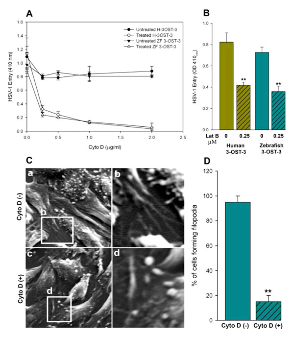
Fig. (3) Actin depolymerizers block HSV-1 entry into zebrafish (ZF) 3-OST-3 expressing CHO-K1 cells. (A, B) Cultured monolayers
of cultured human (H) and zebrafish (ZF) encoded 3-OST-3 CHO-K1 cells were pre-treated with the indicated concentrations of the actin
depolymerizing agent Cytochalisin D (Cyto D; panel A) and Lanticulin B (Lat B; panel B) before exposed to β-galactosidase expressing
HSV-1 gL86 (25 pfu/cell) virus. Cells treated with 1 x PBS treated cells were used as a control. Viral entry was quantitated 6 h after
infection at 410 nm using a spectrophotometer. The experiment was repeated three times with similar results. (C) Visualization on the
inhibition of filopodia via scanning electron microscopy (SEM) on HSV-1 infected (25 pfu/cell for 45 min at 37°C) ZF-3-OST-3 expressing
CHO-K1 cells in absence (panel a) and presence (panel c) of Cyto D at 0.5 µg/ml. The regions in panel a (filopodia induction) and panel c
(reduced filopodia) highlighted are shown in panel b and d. (D) Determination of percentage of filopodia scored in ZF-3-OST-3 infected
cells in absence and presence of Cyto D. Sampled groups of 50 single cells or clusters (with 5-20 cells) in a triplicate experiment, 5 µm
length of a protrusion and at least 10% of the cell surface covered with 25 or more protrusions is scored positive. ** P< 0.05, one way
ANOVA.