- Home
- About Journals
-
Information for Authors/ReviewersEditorial Policies
Publication Fee
Publication Cycle - Process Flowchart
Online Manuscript Submission and Tracking System
Publishing Ethics and Rectitude
Authorship
Author Benefits
Reviewer Guidelines
Guest Editor Guidelines
Peer Review Workflow
Quick Track Option
Copyediting Services
Bentham Open Membership
Bentham Open Advisory Board
Archiving Policies
Fabricating and Stating False Information
Post Publication Discussions and Corrections
Editorial Management
Advertise With Us
Funding Agencies
Rate List
Kudos
General FAQs
Special Fee Waivers and Discounts
- Contact
- Help
- About Us
- Search


The Open Clinical Cancer Journal
(Discontinued)
ISSN: 1874-1894 ― Volume 5, 2011
Psoas Abscess after Radical Abdominal Hysterectomy: A Case Report and Review of the Literature
Dimitrios Spiliopoulos*, Khadra Galaal, Keith Godfrey , Raj Naik
Abstract
We report a case of a 38-year- old woman who presented with right lower quadrant abdominal pain and occasional fevers, five weeks after radical abdominal hysterectomy and pelvic node dissection for cervical carcinoma. After initial empirical antibiotic treatment failure, a decision was made to proceed with percutaneous drainage of a right psoas abscess. The antibiotic regimen was adjusted according to the culture results from the abscess. The patient's condition improved in the next 36 hours. The patient was discharged six days later on oral antibiotic treatment for four weeks.
Psoas abscess is a rare condition in general obstetrics and gynecology and its incidence in gynecological oncology is very low. This is a potentially fatal condition with difficult diagnosis and treatment. Appropriate management of these cases should include suspicion and confirmation of the diagnosis and aggressive medical and surgical treatment.
Article Information
Identifiers and Pagination:
Year: 2010Volume: 4
First Page: 15
Last Page: 19
Publisher Id: TOCCJ-4-15
DOI: 10.2174/1874189401004010015
Article History:
Received Date: 20/5/2010Revision Received Date: 5/8/2010
Acceptance Date: 6/8/2010
Electronic publication date: 29/10/2010
Collection year: 2010
open-access license: This is an open access article licensed under the terms of the Creative Commons Attribution Non-Commercial License (http://creativecommons.org/licenses/by-nc/3.0/) which permits unrestricted, non-commercial use, distribution and reproduction in any medium, provided the work is properly cited.
* Address correspondence to this author at the Gateshead Health NHS Foundation Trust, Northern Gynaecological Oncology Centre, Queen Elizabeth Hospital, Sheriff Hill, Gateshead, NE9 6SX, UK; Tel: (0044) 01914820000; Fax: 004401914456192; E-mail: dimspiliop@yahoo.com
| Open Peer Review Details | |||
|---|---|---|---|
| Manuscript submitted on 20-5-2010 |
Original Manuscript | Psoas Abscess after Radical Abdominal Hysterectomy: A Case Report and Review of the Literature | |
INTRODUCTION
First described by Minter in 1881, psoas abscesses can develop from hematogenous or lymphatic dissemination of an infection from an occult primary source (primary abscess) [1-3]. Direct spread from an adjacent infection of the bowel, the genital or urinary tract and the bone is another mechanism (secondary abscess). It may be uncertain whether invol-vement of an adjacent structure is a cause or a consequence of the abscess [4].
Primary abscesses are more common in tropical and developing countries. In Europe and North America 17 to 61 percent of the abscesses are primary, however, in Asia and Africa 99 percent are primary [3]. In some occasions it can be difficult to differentiate between primary and secondary cause [5].
Risk factors for the formation of primary abscesses are conditions associated with immunosuppression including cancer, diabetes, human immunodeficiency virus (HIV) infection, intravenous drug use, renal failure and chronic steroid use [2,6]. The case described was treated for cervical cancer, a condition that can potentially predispose to suppressed immune status and the development of sepsis in advanced stage.
Psoas abscesses are more common in males than females [3,7] with a median age of 44 to 58 years in developed countries. They occur with the same frequency on the right and left side, bilateral abscesses being uncommon (one to five percent).
Primary psoas abscesses are more frequently caused by a single organism [8]. Staphylococcus aureus has been the most commonly identified microbial organism associated with psoas abscess as it is present in 88% of the cases, toge-ther with Streptococcus (5%) and E. coli (3%). Bacteroides, Enteric pathogens, Streptococcus and Staphylococcus can also be present [3, 9-12]. Mycobacterium tuberculosis is still common in developing countries, but also frequently encountered in HIV infected patients. Secondary psoas abscesses can be monomicrobial or polymicrobial, including enteric organisms.
Psoas abscess is a condition rarely reported in the available obstetric and gynecologic literature. A few cases have been diagnosed after vaginal delivery [13-16], two more were reported during the course of a normal pregnancy [17, 18] and one case was reported after dilation and curretage for abortion with an intrauterine device in place [19].
In gynecological oncology it is very rarely encountered, despite the extensive tissue dissection and degree of radicality involved in many of these procedures. The clinical picture can be variable and nonspecific. It can present with the clinical triad of fever, back pain and groin or thigh pain, although the full triad is present in less than one third of the patients.
We present the first reported case of psoas abscess after radical hysterectomy and pelvic lymph node dissection for treatment of cervical cancer.
CASE PRESENTATION
A 38 year old G4P2 woman diagnosed with a stage 1b1 poorly differentiated adenosquamous cervical carcinoma was treated in our institution with radical hysterectomy and bilateral systematic pelvic lymph node dissection. We performed the lymph node dissection after the uterus was removed. The extent of lymphadenectomy consisted of the removal of all visible pelvic lymph nodes from the bifurcation of the common iliac artery caudally. No enlarged common iliac or paraortic nodes were identified. The fascia of the iliopsoas muscle was incised along the line of the external iliac artery. The incision was carried up to the retractor, and the lateral external iliac nodes lying under the inguinal ligament were excised across the vessels. Small lymphatic channels and blood vessels were clipped with liga clips. The dissection continued along the external iliac artery and a sheet of fascia and nodal tissue was developed leaving the arteries clean from the bifurcation of the common iliac artery to the inguinal ligament. At the upper end of the dis-section, the sheet of tissue was removed across the internal iliac artery. At the final stage, the dissection continued below the external iliac vein, emptying the obturator fossa of all nodes. The block of tissue containing lyphnodes was lifted out. Hemostasis at the end of the operation was meticulous using argon beam coagulation, no pelvic drains were used and the estimated blood loss was 600 ml. The vaginal vault was oversewn and left open, to prevent formation of pelvic lymphocysts or hematomas.
The postoperative recovery was uneventful and the patient was discharged from the hospital seven days postope-ratively. She received a subcutaneous dose (3500IU) of a low molecular weight heparin (tinzaparin) for her entire hospital stay, starting from the afternoon before surgery was performed. A single 1 gram intravenous dose of cefazolin was administered half an hour before the start of the operation. During her postoperative stay, no antibiotic coverage was provided. The pathology report showed no evidence of lymphovascular space involvement, no extension to the parametrial tissues, clear vaginal margins and all lymph nodes (nine on the left and eleven on the right pelvic side) were negative for tumor metastasis. Further treatment with chemotherapy or radiation therapy was not recom-mended.
Five weeks after surgery, she presented to the emergency department complaining of right lower quadrant abdominal pain with occasional fevers for two weeks. The patient was unable to move or flex her right hip and had a swollen right groin. The pain was also radiating to the right leg and required oral morphine.
Her vital signs included a temperature of 39.4 (C, blood pressure of 125/82 mmHg, heart rate of 102 beats per minute and respiratory rate of 13. On physical examination, the patient was alert, awake and oriented to self, time and place. The cardiovascular exam revealed mild tachycardia with regular rhythm. Lungs were clear to auscultation bilaterally. The abdominal examination showed a well-healed midline infraumbilical incision, no evidence of rigidity or rebound tenderness and presence of normal bowel sounds. A tender palpable mass (measuring 5 cm in diameter) was noted in the right iliac fossa. No erythema or echymosis were present on the overlying skin. Pelvic and rectal examination did not reveal any abnormalities. Upon examination of the extremities, flexion of the right hip joint with resistance, abduction and extension were extremely painful with decreased range of motion. Deep tendon reflexes were normal bilaterally. Palpation of the femoral, popliteal and dorsalis pedis arteries revealed normal pulses bilaterally. She had no urinary symptoms.
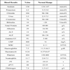
Laboratory investigations included a complete blood count with a White Blood Cell (WBC) count of 14.5 x10^9/L with 81% neutrophils, Hb of 7.5 g/dl, Platelets 404 x10^9/L, Sodium 138 mmol/L, Potassium 4.2 mmol/L, Urea 5.5 mmol/L, Creatinine 74 mmol/L, Albumin 29 g/L, Alanine Transaminase (ALT) 14 u/L, Alkaline Phosphatase (ALP) 185 u/L, Gamma glutamyltransferase (gamma GT) 91 u/L, Prothrombin time (PT) 21 and Partial thromboplastin time (aPTT) 48. Her C-Reactive Protein (CRP) was 468 mg/L (Table 1). Two sets of blood cultures were obtained as well as a urine culture.
Imaging studies included a chest x-ray that was unre-markable, without any effusion or infiltrates. Computed tomography (CT) scan of the abdomen and pelvis demonstrated a large (10 cm diameter) right psoas muscle mass extending along the entire length of the muscle and a large cavity seen in the lower part of the abdomen, with right hydronephrosis and hydroureter. The presence of a well-defined hypodense lesion with an air-fluid level, in the right psoas muscle is evident (Fig. 1 ). The appendix was normal with no sign of inflammation, although loops of small bowel were closely applied to the medial surface of the right psoas mass. Differential diagnosis included infected lymphocyst, hematoma and urinoma. The patient was administered intravenous empirical antibiotic treatment with cefuroxime initially and subsequently amoxycillin. A decision was made to proceed to percutaneous drainage of the abscess by an interventional radiologist. After the positioning of an intraabdominal drain in the radiology department, 100 ml of thick pus were drained and sent for culture, further 600ml of pus was drained over the next 48 hours (Fig. 2
 ). The fever and tachycardia resolved within the next 36 hrs.
 |
Fig. (1) CT image. Well-defined hypo-dense lesion in the right psoas muscle with peripheral enhancement (10 cm). |
 |
Fig. (2) CT image. Same lesion in the right psoas muscle reduced in size after placement of a drain |
The blood cultures obtained on admission were negative as well as the urine culture. The cultures obtained during drainage of the abscess grew E. Coli andHaemolytic streptococcus group C. The antibiotic regimen was adjusted to iv piperacillin/tazobactam and metronidazole according to sensitivity studies. The drain stayed in situ for six days and the patient recovered without further complications. She was discharged home after removal of the catheter on oral coamoxyclav and metronidazole for 4 weeks.
DISCUSSION
Clinical diagnosis of a psoas abscess is challenging since the symptoms and signs can be variable and nonspecific. The majority of patients present with a non-specific history of weight loss, limp, anorexia, inguinal mass, low back or abdominal pain and fever [2, 11, 20-23]. Leukocytosis (>10,000/mL) is observed in up to 83% of cases, elevated CRP and anemia <11 g/L (42.6 % in one series) are quite common. Thrombocytosis is less frequently observed (27 % of cases) [7]. CRP is commonly elevated and on average 189.8 in one series [2]. Aspartate amino-transferase was elevated in 38% of cases (>40) [7].
The symptomatology and blood test results in our case were very similar to those previously described, although the history and physical examination alone can be identical in patients with pelvic abscesses, peritonitis from other causes such as hematoma, lymphocyst or a urinoma. In the case of an infected lymphocyst, this appears a few days after the operation and may grow steadily. On rectal examination, it would be palpable as a smooth, tense mass attached to the pelvic sidewall. Often the patient is febrile in the immediate postoperative period. Our patient did not present with such a symptomatology. Given the reasonably limited blood loss, the good hemostasis at the end of the operation and the open vaginal vault, the presence of a large hematoma was less probable. Also, in our patient, the pelvis was not reperito-nized; significantly reducing the incidence of lymphocyst or hematoma formation.
The main imaging studies, to aid with the diagnosis, are ultrasound and CT scan of the abdomen and pelvis with a sensitivity of 41-95% and 95-100% respectively [11]. The most common CT feature of an abscess is a focal hypodense lesion within a larger muscle. Other specific characteristics are the presence of air-fluid level or gas bubbles and irregular margins of the lesion [24]. CT can also identify a retroperitoneal or intra-abdominal disease process as the cause for the abscess. It also allows the simultaneous drainage of the abscess by percutaneous needle aspiration [11, 25, 26].
Blood cultures and material from the abscess should be sent to the microbiology laboratory for Gram stain and culture. They are positive in 41 to 68 percent of cases; S. Aureus been the most frequent isolate [3, 27]. In our patient, both blood cultures were negative, suggesting a secondary cause, more probably the bowel.
The treatment is based on drainage of the abscess and antibiotics [26]. Secondary abscesses may also require management of the infected focus. The antibiotic treatment should be directed empirically initially against the most common bacteria involved in the process. The appropriate antibiotic treatment should be guided later from blood culture results and culture of the abscess. If there is no signi-ficant improvement with intravenous antibiotic treatment, repeating the culture of the abscess and seeking the advise of microbiologist is a valid option. The drainage of the abscess can be done either under image guidance (CT or ultrasound) or through a laparotomy. Percutaneous drainage is much less invasive and useful in cases of cysts that contain few septa [28]. Antibiotic treatment should be continued for 2 to 3 weeks after improvement of the clinical condition and drainage of the abscess. Only a small number of cases reported were treated successfully with antibiotics alone [29].
In our case, the patient's general condition improved dramatically after the drainage of the abscess. Our patient initially received an empiric course of antibiotics based on the suspected clinical diagnosis. The regimen was adjusted to culture results and sensitivities. We decided to proceed with percutaneous drainage of the abscess secondary to the significant size on CT scan and the persistent fevers despite antibiotic treatment. An open approach with drainage of the abscess in the operating room was avoided as an initial approach, since the patient was septic and had recently undergone laparotomy, increasing the risk of postoperative complications [30].
CONCLUSION
There are few cases of reported psoas abscesses in obstetrics and gynaecology. To our knowledge, this is the first reported case after a gynecological oncology procedure. A psoas abscess should always be suspected after a proce-dure involving retroperitoneal dissection in a patient who presents with the classical triad of fever, abdominal pain and thigh pain on movement. In the majority of the cases however, the general symptomatology will be nonspecific. The clinical suspicion should be higher in lengthy oncologic procedures that include more extensive tissue dissections and thus higher infection rates. Even if the source of infection is not initially evident, the clinician must act quickly and aggressively to prevent further complications.