- Home
- About Journals
-
Information for Authors/ReviewersEditorial Policies
Publication Fee
Publication Cycle - Process Flowchart
Online Manuscript Submission and Tracking System
Publishing Ethics and Rectitude
Authorship
Author Benefits
Reviewer Guidelines
Guest Editor Guidelines
Peer Review Workflow
Quick Track Option
Copyediting Services
Bentham Open Membership
Bentham Open Advisory Board
Archiving Policies
Fabricating and Stating False Information
Post Publication Discussions and Corrections
Editorial Management
Advertise With Us
Funding Agencies
Rate List
Kudos
General FAQs
Special Fee Waivers and Discounts
- Contact
- Help
- About Us
- Search


The Open Anatomy Journal
(Discontinued)
ISSN: 1877-6094 ― Volume 6, 2014
Anomaly of the Sternothyroideus Muscle Insertion in a Dog
Maria Isabel Giner, Begoña Ballester-Lurbe, Olga Gomez , Jose Terrado*
Abstract
In this work, we describe an anatomic anomaly of the sternothyroideus muscle for the first time. The dissection of the left side of the neck of a young adult female dog showed a group of fibres of the sternothyroideus muscle diverging cranially and ventrally from the caudal region of the muscle onwards to be inserted at the thyrohyoideum bone, close to the insertion site of the sternohyoideus muscle. Dissection of the right aspect of the neck revealed a normal right sternothyroideus muscle. The consequence of the presence of this muscle strip was a link between the sternothyroideus muscle and the apparatus hyoideus, and it can be interpreted as a sign of the close phylogenetic relationship between the sternothyroideus and the sternohyoideus muscles.
Article Information
Identifiers and Pagination:
Year: 2009Volume: 1
First Page: 11
Last Page: 12
Publisher Id: TOANATJ-1-11
DOI: 10.2174/1877609400901010011
Article History:
Received Date: 3/7/2009Revision Received Date: 12/8/2009
Acceptance Date: 10/9/2009
Electronic publication date: 05/11/2009
Collection year: 2009
open-access license: This is an open access article licensed under the terms of the Creative Commons Attribution Non-Commercial License (http: //creativecommons.org/licenses/by-nc/3.0/ which permits unrestricted, non-commercial use, distribution and reproduction in any medium, provided the work is properly cited.
* Address correspondence to this author at the Department of Animal Medicine and Surgery, Faculty of Veterinary Science, CEU-Cardenal Herrera University, Avda Seminario, s/n 46113 Moncada-Valencia, Spain; Tel: ++ 34 96 1369000; Fax: ++ 34 96 1395272; E-mail: jterrado@uch.ceu.es
| Open Peer Review Details | |||
|---|---|---|---|
| Manuscript submitted on 3-7-2009 |
Original Manuscript | Anomaly of the Sternothyroideus Muscle Insertion in a Dog | |
The ventral neck region of the dog comprises a group of muscles which are intimately related with the trachea and oesophagus. The main muscles covering this area ventrally and laterally are the brachiocephalicus, sternocephalicus, sternohyoideus and sternothyroideus. The brachiocephalicus and sternocephalicus are superficially and laterally found. The sternohyoideus is medially located in the ventral midline of the neck covering the ventral surface of the trachea. The sternothyroideus muscle is closely related to the sternohyoideus. It emerges from the first costal cartilage to be inserted at the lateral surface of the thyroid cartilage of the larynx. It is deeply located at the sternocephalicus and the sternohyoideus muscles and its medial surface is in contact with the trachea. The sternothyroideus is a long flat muscle with a small tendinous transverse intersection. Its main action consists in pulling the larynx and, subsequently, the apparatus hyoideus and tongue caudally [1].
Twelve mongrel dogs were routinely prepared by vascular perfusion of fixative solutions to be studied in the Anatomy and Embryology course at the Faculty of Veterinary Sciences (University CEU-Cardenal Herrera, Moncada, Valencia, Spain) during the academic year 2007-2008. Cadavers were obtained from a local shelter, while the authorisation for the procedure was obtained from both the Local Authorities and the University’s Ethical Committee. After fixation, animals were anatomically dissected. We describe here an anomaly found in one young adult female dog.
The dissection of the left side of the neck in one young adult female mongrel showed a group of fibres moving away from the sternothyroideus muscle. This group of fibres diverged from the caudal region of the muscle and adopted a new slightly more ventrally position to the normal muscle (Fig. 1A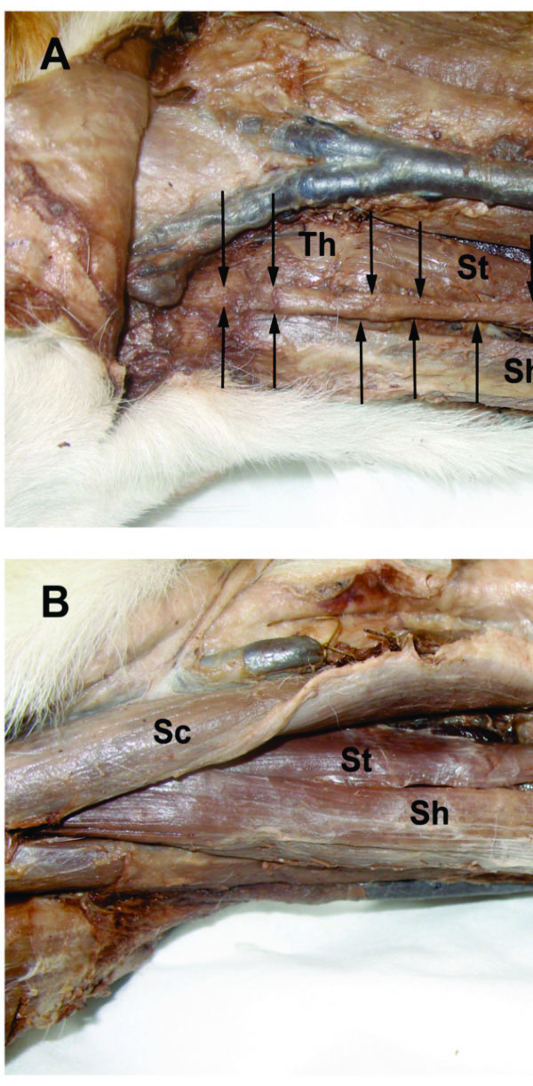 ). This anomalous strip was inserted at the ventral part of the thyrohyoid bone, ventrally to the thyrohyoideus muscle insertion and dorsolaterally to the insertion of the sternohyoideus muscle at the basihyoid bone. Thus, this band had unique features of origin and insertion which were not present in either the normal sternothyroideus or strenohyoideus muscles. Apart from the anomalous band, the sternothyroideus muscle was, as usually described, inserted at the thyroid cartilage of the larynx. Whereas the sternothyroideus muscle was 16.5 cm long and 1.3 cm wide, the anomalous band was 11 cm long and 0.5 cm wide. Although it was not possible to perform histological sections, a macroscopic analysis of the muscle (including the anomalous slip) revealed no apparent deformity of the muscle fibres or in the conjunctive tissue. Dissection of the right aspect of the neck showed the right sternothyroideus muscle in its standard position (Fig. 1B), and it was inserted normally at the lateral surface of the thyroid lamina. Neither the right nor the left sternohyoideus muscles showed any apparent anomaly and no other muscular anomaly was detected in this dog.

Although muscular anomalies are sporadically found in domestic mammals, few have been reported in the literature. However it seems that the muscles at the ventrolateral part of the dog’s neck are particularly susceptible to undergoing variations in their attachments. Anatomical anomalies affecting hyoid muscles in dogs were described by Evans [2] who showed variations in the insertions in the digastricus, stylohyoideus, milohyoideus and sternohyoideus muscles in Beagles and mongrels. A wider variation was observed in Beagles and was considered a specific breed feature. More recently, we have observed the absence of the left thyrohyoideus muscle in a dog [3], and slips of the sternohyoideus similar to those previously reported by Evans (not shown). Of a total of 160 dogs dissected over a ten-year period, we have not previously observed the abnormality reported herein, and to our knowledge this is the first description showing an anomaly affecting the sternothyroideus muscle. However, variations such as the doubling, absence or accessory slips to the thyrohyoideus, the inferior constrictor or the carotid sheath have been reported in humans [4]. The band of the sternothyroideus muscle herein described in a dog could be considered an “intermediate muscle” between the sternothyroideus and the sternohyoideus muscles since it runs from the caudal part of the former to be closely inserted to the latter. The sternohyoideus muscle pulls the hyoid apparatus and, subsequently, the tongue caudally. The sternothyroideus draws the larynx caudally, but it can also indirectly collaborate with the sternohyoideus function to pull the hyoid apparatus caudally through the thyrohyoideus muscle and the thyrohyoid membrane which connect the thyroid cartilage and the hyoid apparatus. It is conceivable that the anomalous slip could help in these functions.
Hyoid muscle anomalies have been explained in the light of phylogeny on the basis of differential development and muscle migration, which results in muscle patterns that are reminiscent of “lower” animals [2]. A phylogenetic explanation could also be applied to an abnormally caudal insertion of the splenius muscle reported in a dog [5]. In this case, the anomaly is reminiscent of the presence of the splenius cervicis muscle, which is absent in dogs but present in other mammals. Conversely, the anomalous slip of the sternothyroideus that we present herein is not present, to our knowledge, in any mammalian species. The sternothyroideus is closely related, physically and phylogenetically, to the sternohyoideus muscle, and even these are mixed in some species like the Ornithorhyncus anatinus or the Tupaia sp [6, 7]. Both muscles belong to the group of hypobranchial muscles, which originate from the cervical somites that migrate ventrally. The plesiomorphic condition of hypobranchial muscles for sarcopterigians, including tetrapods, is thought to be composed of two muscles that are mainly related to the opening of the mouth, the coracomandibularis and the sternohyoideus. While the sternohyoideus is undivided in fishes, amphibians and reptiles, two new muscles, the sternothyroideus and the thyrohyoideus, are considered to derive from it in mammalian species [6, 8]. The band that we have observed, as well as those described by Evans (both representing “anomalies” which, in some way, connect the sternothyroideus and the sternohyoideus), are probably signs of this common origin of these muscles. Nevertheless, this type of anomalies can also be considered fortuitous variations, probably due to small malformations by altered migrations of forming cells during the muscular system development.
ACKNOWLEDGEMENTS
This work has been supported by PRCEU-UCH grants from the CEU-Cardenal Herrera University and from the Copernicus–Santander Research Programme. Fellowship support for B. Ballester-Lurbe from the Generalitat Valenciana is also gratefully acknowledged.