- Home
- About Journals
-
Information for Authors/ReviewersEditorial Policies
Publication Fee
Publication Cycle - Process Flowchart
Online Manuscript Submission and Tracking System
Publishing Ethics and Rectitude
Authorship
Author Benefits
Reviewer Guidelines
Guest Editor Guidelines
Peer Review Workflow
Quick Track Option
Copyediting Services
Bentham Open Membership
Bentham Open Advisory Board
Archiving Policies
Fabricating and Stating False Information
Post Publication Discussions and Corrections
Editorial Management
Advertise With Us
Funding Agencies
Rate List
Kudos
General FAQs
Special Fee Waivers and Discounts
- Contact
- Help
- About Us
- Search


The Open Anatomy Journal
(Discontinued)
ISSN: 1877-6094 ― Volume 6, 2014
Argyrophil Cells in Normal Human Uterine Cervix
Sujatha D’Costa*, 1, C.V Raghuveer2, Adhikari Prabha3, Sampath Madhyasta1
Abstract
Introduction:
Argyrophil cells are neuroendocrine cells which react with silver stain resulting in brown or black coloration with the help of an external reducer. Argyrophil cells occur rarely in the normal human uterine cervix but are found in a variety of invasive tumors arising in the cervix.
Methods and Results:
About 317 normal human uterine cervices were stained with Grimelius argyrophilic reaction out of which 158 cases (49.84%) showed the positivity. The immunomarkers used in the present study were Chromogranin A and Synaptophysin.
Discussion:
In the present study, an attempt has been done to study the role of Argyrophil cells in normal uterine cervix using a larger sample size. The earlier studies have shown that the carcinoid tumors of the uterine cervix may a have arisen from argyrophil cells of the normal uterine cervix and are part of the group of neoplasms of the diffuse endocrine cell system (Amine Precursor Uptake and Decarboxylation or APUD). Detection and evaluation of argyrophil cells is important in assessment of the progression of precancerous lesions of cervix.
Article Information
Identifiers and Pagination:
Year: 2010Volume: 2
First Page: 25
Last Page: 28
Publisher Id: TOANATJ-2-25
DOI: 10.2174/1877609401002010025
Article History:
Received Date: 3/9/2009Revision Received Date: 29/10/2009
Acceptance Date: 19/11/2009
Electronic publication date: 31/3/2010
Collection year: 2010
open-access license: This is an open access article licensed under the terms of the Creative Commons Attribution Non-Commercial License (http://creativecommons.org/licenses/by-nc/3.0/) which permits unrestricted, non-commercial use, distribution and reproduction in any medium, provided the work is properly cited.
* Address correspondence to this author at the Department of Anatomy, Centre for Basic Sciences, Kasturba Medical College, Bejai, Mangalore, PIN 575004, Karnataka, India; Tel: +91 824 2222768; Fax: +91 824 2428183; E-mails: sujatha.dcosta@manipal.edu; sujathasn@gmail.com
| Open Peer Review Details | |||
|---|---|---|---|
| Manuscript submitted on 3-9-2009 |
Original Manuscript | Argyrophil Cells in Normal Human Uterine Cervix | |
INTRODUCTION
The transformation zone of the uterine cervix is of utmost importance because cervical cancer and its precursors typically begin within it [1, 2]. Argyrophil cells occur rarely in the normal human uterine cervix but are found in a variety of invasive tumors arising in the cervix. The Grimelius technique shows that the positive cells are usually triangular or flask shaped with broad base in the opposition to a basement membrane of the epithelium [3, 4] prolonged by one or two cytoplasmic spurs extending from the basal membrane towards the lumen, which the spurs rarely reached. According to Fox et al. [5] argyrophil cells have been demonstrated in the GIT (Gastro Intestinal Tract), the pancreas, the biliary tract, the adrenal medulla, the organ of Zuckerkandl, the carotid body and the prostate. Argyrophil cells are not present in normal human ovary, fallopian tube or endometrium, but occasionally they are found in the normal cervix. Scanty argyrophil cells are present in a substantial proportion of normal endometrial, particularly during the secretory stage of the cycle. Presence of neuroendocrine cells in some pathological conditions including cervical carcinomas has been detected by Fetissof et al. [6]. In a primary carcinoid tumor of the uterine cervix, Albores – Saavedra, Poucell & Rodriguez [7] reported the presence of argyrophilic cells, although, without any information on qualitative and quantitative parameters. However, these authors have provided the data to support that, these cells are found among the linings of endocervical glands and in cervical squamous epithelium of uterine cervix. Isolated neuroendocrine epithelial cells of argentaffin type or argyrophil type are admixed with the normal endocervical cells. Argyrophil cells need the assistance of an external reducing agent to reduce silver solution to metallic silver (in vivo) [8]. Cells and tissues which require the use of extraneous reducers are termed argyrophil [9, 10]. They ectopically produce a variety of amines and peptide hormones. Argyrophil cells are suggested to be a counterpart of the neoplasm (APUDoma) derived from APUD (Amine Precursor Uptake and Decarboxylation) cells [11].
A systemic detection of argyrophil cells in the normal uterine cervix was carried out, using Grimelius argyrophilic reaction and immunohistochemical techniques.
MATERIALS AND METHODS
Around 317 paraffin embedded blocks of uterine cervices were studied from hysterectomy specimens which had their histopathological diagnosis confirmed in the Pathology department K.M.C. Mangalore. The study was approved by the Ethical Committee. Several coronal sections of 4 to 5 μ thickness were cut from the above paraffin blocks and were transferred to the slide. One section from each paraffin block were deparaffinised and stained with Hematoxylin and Eosin (H&E) using standard procedure [12]. The sections were examined to confirm the presence of stratified squamous keratinized epithelium (Fig. 1A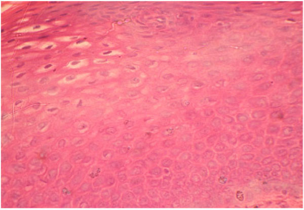 , B
, B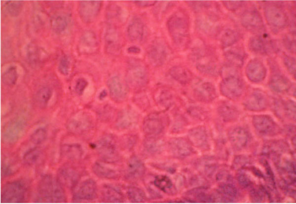 ). The unstained sections of the same series were stained with Grimelius Argyrophilic reaction as per standard procedure. The Grimelius argyrophilic reaction is a well known procedure was used by employing the principle of impregnation with silver nitrate (AgNO3) and reduction with hydroquinone and sodium sulphate. The Grimelius argyrophilic reaction is useful in detecting highly or moderately granulated neoplasms [13].
 |
Fig. (1A) Haematoxylin & Eosin staining at 40X. |
 |
Fig. (1B) Haematoxylin & Eosin staining at 100X. |
Tissue known to contain argyrophil granules such as spinal ganglion was used as a control for the Grimelius argyrophilic reaction.
The sections which showed positivity with Grimelius argyrophilic reaction were selected. The unstained sections of the same series were selected and used for immunohistochemistry method using Chromogranin A and Synaptophysin as argyrophilic cell markers. Positive results aid in the classification of neuroendocrine tumours.
RESULTS
In our study 317 normal uterine cervices were stained with Grimelius argyrophilic reaction to evaluate the presence of argyrophilic cells. Out of which 158 cases (49.8%) were positive. The Argyrophil granules of the endocrine cells were impregnated yellow brown to black (Fig. 1C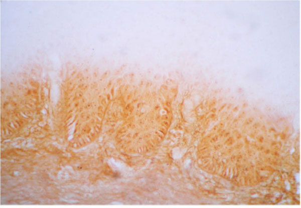 , D
, D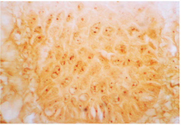 ).
).
 |
Fig. (1C) Grimelius` Argyrophil Reaction at 40X. |
 |
Fig. (1D) Grimelius` Argyrophil Reaction at 100X. Arrows indicate the Argyrophil cells. |
About 60 normal uterine cervices were used in Immunohistochemistry method for Chromogranin A, out of which 28 were positive (46.6%) (Fig. 1E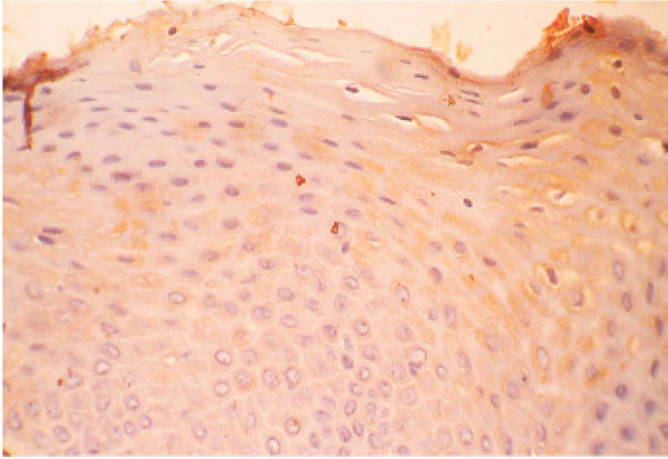 , F
, F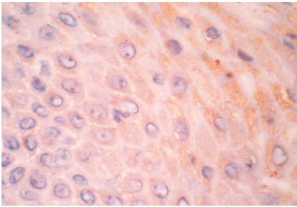 ) and 26 (43.3%) were positive with Synaptophysin marker (Fig. 1G
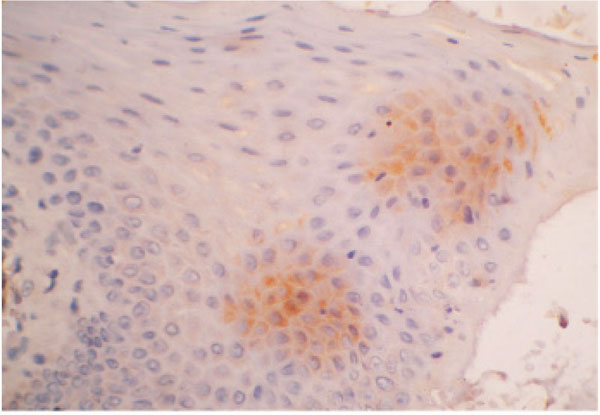 , H
, H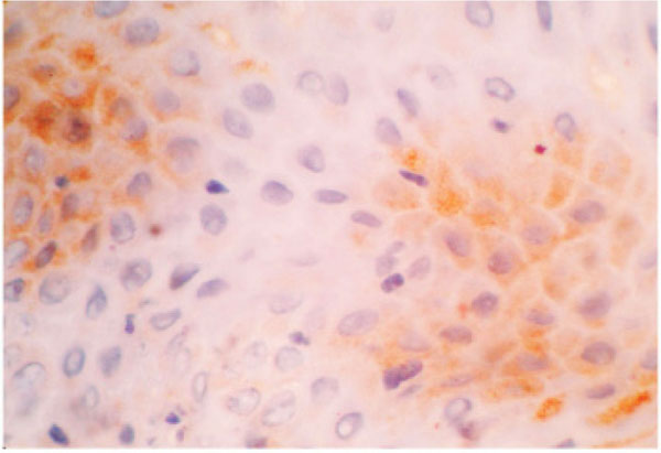 ). The argyrophilic granules in the positive cells were immunostained into golden color.
 |
Fig. (1E) Immunoexpression with Chromogranin A at 40X. |
 |
Fig. (1F) Immunoexpression with Chromogranin A at 100X. Arrows indicate the Cg A reaction. Location of reaction: Cytoplasmic. Intensity of reaction: Weak. |
 |
Fig. (1G) Immunoexpression with Synaptophysin at 40X. |
 |
Fig. (1H) Immunoexpression with Synaptophysin at 100X. Arrows indicate the SYN reaction. Location of reaction: Cytoplasmic. Intensity of reaction: Moderate. |
DISCUSSION
Fox et al. [5] demonstrated the presence of argyrophilic cells in 2 out of 120 normal cervices (1.6%). Tateishi et al. [14] were also able to demonstrate argyrophilic cells among normal cervical epithelium in 19 out of 54 patients (35.18%). Fetissof et al. [15] identified argyrophilic cells in 11 out of 210 ectocervices (5.2%). Argyrophil cells have been documented in the normal uterine cervix by several authors [16-18]. Fetissof et al. [19] found two types of argyrophilic cells in 43% of ectocervices: serotonin cells and Merkel - type cells.
Our study scores over these studies in view of the larger sample size and higher percentage of positive cases (nearly 50%).
In the present study, we have studied for chromogranin A and Synaptophysin (SYN) positivity in all cases that showed argyrophilia. Neuroendocrine tumors of the uterine cervix describe cervical neoplasms that show the histological characteristics of carcinoids, including argyrophilia and/or argentaffinnia, and immunoreactivity for chromogranin, synaptophysin, and neuron-specific enolase [7]. Chromogranin A is considered the best general neuroendocrine serum or plasma marker available both for diagnosis and therapeutic evaluation and is increased in 50-100% of patients with various neuroendocrine tumors. Chromogranin A serum or plasma levels reflect tumor load, and it may be an independent marker of prognosis in patients with midgut carcinoids [20]. According to Luca et al. [21] the serum Chromogranin A increases in patients with neuroendocrine derived neoplasms.
The neuroendocrine marker SYN is expressed in a small subset of non-small-cell carcinomas of the cervix and its expression seems to correlate with a poor outcome [22]. Immunohistochemical studies performed in 23 primary small cell carcinoma of the uterine cervix showed immunoreactivity for chromogranin in 10 tumors (43%), 13 for synaptophysin (56%) [23].
Six cases of cervical large cell neuroendocrine carcinomas (LCNEC) out of 972 were immunoreactive for chromogranin A and /or synaptophysin. These observations provide potential support for the derivation of LCNECs from the endocrine argyrophil cells in the normal cervix [24]. The large majority of these cervical neoplasms are histologically and clinically aggressive [25]. Consequently, the search for Chromogranin A and Synaptophysin markers is useful in identifying neuroendocrine cells. The functions of the neuroendocrine cells are related to the synthesis and secretion of numerous hormonal peptides, amines and growth factors, which make use of endocrine, paracrine and autocrine mechanisms to regulate a wide variety of biological functions [21]. Immunohistochemical analysis using several kinds of neuroendocrine markers is helpful in establishing the correct diagnosis in addition to focusing on characteristic histo- and cytopathologic features [26].
In the present study 317 normal uterine cervices were stained with Grimelius argyrophilic reaction to evaluate the presence of argyrophilic cells. Out of which 158 cases (49.84%) were positive. About 60 normal uterine cervices were used in Immunohistochemistry method for Chromogranin A, out of which 28 (46.6%) and for Synaptophysin 26 (43.3%) were positive.
To our knowledge, this is the first report in view of the larger sample size and higher percentage of positive cases of argyrophilia in the normal uterine cervix. The earlier studies have shown that the carcinoid tumors of the uterine cervix may a have arisen from argyrophil cells of the normal uterine cervix and are part of the group of neoplasms of the diffuse endocrine cell system (APUD). Study of argyrophilia may be of importance to compare the occurrence in normal cervix and compare with the different lesions of the uterine cervix which may help in prognostication.
ACKNOWLEDGEMENT
The authors are indebted to the faculty of Pathology Department, Kasturba Medical College Mangalore, for providing the hysterectomy specimens with histopathological diagnosis.