- Home
- About Journals
-
Information for Authors/ReviewersEditorial Policies
Publication Fee
Publication Cycle - Process Flowchart
Online Manuscript Submission and Tracking System
Publishing Ethics and Rectitude
Authorship
Author Benefits
Reviewer Guidelines
Guest Editor Guidelines
Peer Review Workflow
Quick Track Option
Copyediting Services
Bentham Open Membership
Bentham Open Advisory Board
Archiving Policies
Fabricating and Stating False Information
Post Publication Discussions and Corrections
Editorial Management
Advertise With Us
Funding Agencies
Rate List
Kudos
General FAQs
Special Fee Waivers and Discounts
- Contact
- Help
- About Us
- Search


The Open Arthritis Journal
(Discontinued)
ISSN: 1876-5394 ― Volume 7, 2014
Low-Field Magnetic Resonance Imaging of the Thumb Base in Severe Symptomatic Osteoarthritis: A Comparison with Radiographs and Clinical Symptoms
Gudmundur J. Eliasson*, 1, Eythor Bjorgvinsson2 , Helgi Jonsson*, 3
Abstract
The objectives of this pilot study were to assess the findings of low-field MRI in symptomatic thumb base osteoarthritis (OA), to identify suitable MRI sequences with or without Gadolinium contrast, and to compare these findings with conventional radiographs scored by the Altman atlas and patient reported pain.
Twelve patients with symptomatic thumb base osteoarthritis participated. We used a 0,2T MRI system, before and after i.v. Gadolinium. The MRI images were scored on a 0-3 scale for synovitis (SYN), bone marrow edema (BME), cysts/erosions (CE) and osteophytes (OP).
Interreader agreement on MRI scores was best for BME and CE but less for SYN and OP. Difficulties were in determining the soft tissue involvement of the CMC1 joint, especially in demarcating the joint capsule and ligament pathology. The most informative MRI sequences were the High Resolution Gradient Echo, the STIR and the 3D volume.Gadolinium contrast provided little additional information.
Cumulative MRI and radiographic scores for the 24 CMC1 joints studied were strongly correlated (Rs 0.81, p<0.001). Both BME in adjacent bones and SYN in the CMC1 joint correlated with rest pain (Rs 0.54, p<0.01 and Rs 0.45, p<0.05) respectively). BME was also associated with pain intensity on a 0-10 scale (R 0.44, p<0.05). Other radiographic and MRI variables showed no association with pain.
Low-field MRI reflects CMC1 joint osteoarthritis damage and gives additional information regarding bone marrow edema and synovitis, both of which are strongly related to clinical symptoms. This study encourages further research to identify the most suitable equipment and sequences and to standardize the reading of MRI images.
Article Information
Identifiers and Pagination:
Year: 2009Volume: 2
First Page: 1
Last Page: 6
Publisher Id: TOARTHJ-2-1
DOI: 10.2174/1876539400902010001
Article History:
Received Date: 15/9/2008Revision Received Date: 19/12/2008
Acceptance Date: 23/12/2008
Electronic publication date: 21/1/2009
Collection year: 2009
open-access license: This is an open access article licensed under the terms of the Creative Commons Attribution Non-Commercial License (http: //creativecommons.org/licenses/by-nc/3.0/ which permits unrestricted, non-commercial use, distribution and reproduction in any medium, provided the work is properly cited.
* Address correspondence to these authors at the Roentgen Domus Medica, Egilsgata 3, IS-101 Reykjavik, Iceland; E-mail: gje@simnet.is Department of Rheumatology, Landspitalinn University Hospital, IS-108 Reykjavik, Iceland; E-mail: helgijon@landspitali.is
| Open Peer Review Details | |||
|---|---|---|---|
| Manuscript submitted on 15-9-2008 |
Original Manuscript | Low-Field Magnetic Resonance Imaging of the Thumb Base in Severe Symptomatic Osteoarthritis: A Comparison with Radiographs and Clinical Symptoms | |
BACKGROUND
Osteoarthritis (OA) of the thumb base is a common and disabling form of osteoarthritis [1-3]. It is characterized by a slow variable course and sometimes by episodes with severe symptoms. Standard radiographs are considered the gold standard for the diagnosis of this condition [4], but they have serious limitations, mainly by not reflecting disease activity and thus the relation between radiologic changes and symptoms is generally poor [5]. Furthermore, the slow radiographic progression of OA hampers therapeutic studies which have to go on for years to register effects on disease progression. More dynamic imaging is needed to reflect the ongoing disease progress and both scintigraphy and ultrasound have been advocated in this respect [6, 7].
Magnetic resonance imaging (MRI) has been extensively studied in knee OA. It has several advantages over radiographs in the diagnosis and understanding the disease process. Better imaging of soft tissues such as cartilage, ligaments and joint capsule along with inflammatory and reactive changes in the joints and bones has wastly improved our understanding of the disease process. “Bone marrow edema” (BME) in particular has been found to associate with symptoms and progression of knee OA [8].
MRI imaging of the hands in rheumatoid arthritis is now gaining acceptance as a valuable tool to assess prognosis and monitor the disease progress [9, 10]. We are not aware of previous studies using MRI in thumb base OA.
The disadvantages of MRI imaging include cost, availability, long examination times and most of all, difficulty in reading and interpretation of the findings. We report here a pilot study aiming at determining the most suitable low-field MRI sequences for the assessment of thumb base OA comparing them with symptoms and conventional radiographs. The MRI equipment chosen was an office-based 0,2T system with and without the use of gadolinium contrast.
PATIENTS AND METHODS
Twelve consecutive hand OA patients presenting with clinical symptomatic thumb base osteoarthritis based on squaring and suluxation of that joint in the absence of other rheumatological diagnoses were included in the study. An additional requirement was that the symptoms were predominantly unilateral and the reported pain in the symptomatic joint for the last 48 hours had to be at least 5 on an 0-10 scale. After informed consent, they registered pain on a 0-10 scale in both thumb bases and reported on the presence or absence of rest pain. There were 11 females and 1 male, median age was 57, range 45-71. One of the patients developed a contralateral flare in symptoms while waiting for MRI and thus reported equal pain on both sides. Rest pain was reported by 8 patients, unilateral in all cases.
All patients had standard digital radiographs of the hands. They were scored by both radiologists (GJE,EB) according to the Altman atlas [4], scoring osteophytes (0-3), joint space narrowing (0-3) and erosions (0-3). All readings of radiographs were performed blindly.
MRI imaging was done by a 0,2T Artoscan-Dedicated MRI system from ESAOTE, Italy. The coil used was a dual phased array wrist coil. The MRI sequences were all in the coronal plane; High Resolution Gradient Echo (HRGE) (TR580, TE16), Gradient Echo STIR (TR1120, TE16), Turbo 3D T1 (TR40, TE16) and Spin Echo T1 (TR600, TE26). The field of view was 12cm, interspace 2mm, section thickness 3mm for all sequences except 0.6mm for 3D T1. The imaging display matrix was 512 × 512 for HRGE and 256 × 256 for the other sequences The more symptomatic hand had T1 sequences before and after 10ml i.v. Gadolinium.
The MRI images of the CMC1 joints were scored on a 0-3 scale for synovitis (SYN) (based on capsular hypertrophy and joint effusion), bone marrow edema (BME), cysts/erosions (CE) and osteophytes (OP). Due to difficulties in measuring SYN quantitatively, an additional measure of right-left comparison was added. Consensus scores for both radiographs and MRI images were reached at a second sitting.
The study was approved by the Icelandic National Ethics Committee.
Statistics
All 24 CMC1 joints were used for statistical calculations, comparing the joint with predominant symptoms with the less symptomatic side. The intra-class coefficient (ICC) was used to assess interreader agreement., the Spearman rank test (Rs) for correlation and chi-square for comparison between groups. Multivariate analysis was done by backwards linear regression.
RESULTS
Interreader agreement on MRI scores of the CMC1 joint varied considerably and was acceptable for BME (ICC 0.88) and CE (ICC 0.88) but less so for SYN (ICC 0.63) and OP (ICC 0.66). In the case of SYN, all 24 joints examined had some degree of synovial reactive changes, but there were reading difficulties in determining the soft tissue involvement of the CMC1 joint, especially demarcating the joint capsule and ligament pathology.
For best anatomical-pathological visualisation we found the most informative MRI sequences in this study to be the HRGE, STIR and the 3D T1 volume. HRGE has the best resolution, STIR is essential to demonstrate bone marrow edema and the 3D T1 volume allows evaluations in all planes and visualisation of smaller structures such as osteophytes and cysts. Ordinary dose of Gadolinium contrast provided little additional information. Chosen examples of MRI sequences from two joints are shown below (Figs. 1 and2
and2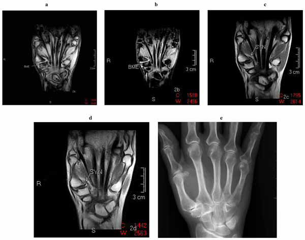 ).
).
 |
Fig. (1) Examples of thumb base MRI images. a) HRGE b) STIR c) T1 before gadolinium d) T1 after gadolinium e) Digital radiograph. |
 |
Fig. (2) Examples of thumb base MRI images. a) HRGE b) STIR c) T1 before gadolinium d) T1 after gadolinium e) Digital radiograph. |
On comparison between MRI and radiographic scores, there was little correlation between the four MRI variables and the three variables scored on the radiographs. On the other hand, aggregate MRI scores showed a very high degree of correlation with aggregate radiography scores for the CMC1 joint (Rs 0.81, p<0.001)(Fig. 3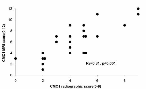 ). The aggregate scores did not show any significant association with symptoms. A summary of radiographic and MRI scores is given in Table 1.
 |
Fig. (3) The relationship between of aggregate CMC1 joint scores from radiographs and MRI. |

On bivariate analysis of the relation between MRI imaging and symptoms, both BME (Rs 0.54, p<0.01) (Fig. 4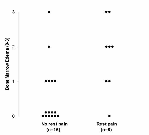 ) and SYN (Rs 0.45, p<0.05) correlated significantly with rest pain and BME also correlated with pain intensity (Rs 0.44, p<0.05) (Fig. 5
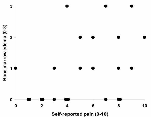 ). The only patient who had a BME score of 3 in the absence of rest pain had more BME on the other side and reported rest pain only on that side. Odds ratios for the co-occurrence of rest pain and BME were clearly significant even with different cut-off points for BME. (BME>1 vs. rest pain; OR 11.7 (1.1-120), p<0.05, BME≥2 vs. rest pain: OR 11.7 (1.5-92), p<0.05). On multivariate analysis however, the independent effect of SYN on rest pain did not reach significance due to its association with BME. However, ten of the eleven patients who reported more symptoms on one side had more SYN on the painful side (p<0.01) although it did not register as a difference on the 0-3 scale. Neither CE nor OP showed a significant relation to pain nor did any of the radiographic scores.
 |
Fig. (4) Patient reported rest pain in relation to bone marrow edema. |
 |
Fig. (5) Patient reported pain severity in relation to bone marrow edema (Rs 0,44, p<0.05). |
DISCUSSION
In this pilot study of 24 CMC1 joints in 12 patients with severe symptomatic thumb base osteoarthritis, scores from MRI images showed a significant association with patient reported pain. No such association was seen with conventional radiographs. The presence of bone marrow edema in particular showed a clear relation to both rest pain and pain severity. Synovitis was also associated with symptoms, but quantitation of synovitis proved problematic with our equipment, despite the use of gadolinium contrast.
One of our aims was to determine the most useful low-field MRI sequences, thus optimizing the use of the equipment and shortening examination times. The ideal situation for clinical use would be having a non-invasive standard run of a few chosen sequences without contrast and a rapid reading protocol. We had planned to extend this study, investigating a larger group of patients with a modified protocol, using only selected sequences. However, the problems of quantifying synovitis indicated a need for a different protocol. Doubling the dose of gadolinium contrast has been shown to improve clarity of synovial tissues [11], but this increases the cost of the examination considerably. Instead, we intend to use higher resolution MRI equipment aiming for increased resolution of soft tissues and cartilage, preferably without the use of contrast to minimize patient discomfort and to shorten the time of aquisition.
Severe symptomatic flares of thumb base OA are fairly common in the clinical situation. Despite that, optimal treatment is in dispute. Intraarticular glucocorticoid injections are widely used and recommended in the European League against Rheumatism (EULAR) guidelines for the treatment of hand OA [12]. This is in spite of a recent placebo controlled study showing no effect of such treatment [13]. In an interesting study of which MRI findings predicted a favorable effect of glucocorticoid injections in the acromioclavicular joint, the presence of synovitis was the best predictor of response [14]. It is easy to postulate that the information from MRI imaging may affect the therepeutic approach to symptomatic thumb base OA; synovitis may well benefit from local injections, whereas bone marrow edema could require more bone directed treatment, local or systemic. We are also in possession of data indicating that MRI images are useful in studies of such therapeutic interventions [15].
The main limitations of this study was naturally its small size and the use of statistical methods on bilateral joints. The statistical results have therefore to be interpreted with great caution.
Finally, we conclude that low-field MRI is a very promising method for the assessment of thumb base OA. The information regarding bone marrow edema and synovitis adds a new dimension to the understanding of pain in the CMC1 joints, particularly as these conditions may warrant different therapeutic approaches. This study encourages further research to identify the most suitable equipment and sequences and to standardize the reading of the MRI images.
ACKNOWLEDGEMENTS
This work was supported by Roentgen Domus Medica, the University of Iceland Research Fund and the Icelandic Osteoarthritis Research Fund.
Thanks to: Our participants for their patience and to radiographers Gudrun Fridriksdottir and Thuridur Halldorsdottir.