- Home
- About Journals
-
Information for Authors/ReviewersEditorial Policies
Publication Fee
Publication Cycle - Process Flowchart
Online Manuscript Submission and Tracking System
Publishing Ethics and Rectitude
Authorship
Author Benefits
Reviewer Guidelines
Guest Editor Guidelines
Peer Review Workflow
Quick Track Option
Copyediting Services
Bentham Open Membership
Bentham Open Advisory Board
Archiving Policies
Fabricating and Stating False Information
Post Publication Discussions and Corrections
Editorial Management
Advertise With Us
Funding Agencies
Rate List
Kudos
General FAQs
Special Fee Waivers and Discounts
- Contact
- Help
- About Us
- Search


The Open Arthritis Journal
(Discontinued)
ISSN: 1876-5394 ― Volume 7, 2014
Antigen-Presenting Cells and their Fcγ and Toll-Like Receptors: Leading Suspects in Autoimmunity
Kim C.M. Santegoets, Lenny van Bon, Mark H. Wenink*, Wim B. van den Berg
Abstract
Antigen-presenting cells (APCs) play an important role in the development of autoimmune diseases. These cells recognize pathogen associated molecular patterns but also endogenously produced ligands through toll-like receptors (TLRs). Aberrant activation of these receptors and the following intracellular signaling pathways can induce the deleterious production of pro-inflammatory cytokines. In genetically predisposed individuals this might lead to a breach in tolerance and eventually autoimmunity. IgG and IgG immune complexes (ICs), which are abundantly present in autoimmune diseases like systemic lupus erythematosus (SLE), rheumatoid arthritis (RA) and systemic sclerosis (SSc) are recognized by APCs via Fc gamma receptors (FcγRs) and can also modulate their activation state. Upon their uptake specific antigens present in ICs are capable of stimulating APCs via their intracellular TLRs, increasing their capability to induce (autoreactive) T and B cell responses. This underscores their likely role in the generation and maintenance of autoimmunity. By focusing on three autoimmune diseases, SLE, RA and SSc, we will illustrate the importance of TLRs and FcγRs in the pathogenesis of autoimmune diseases.
Article Information
Identifiers and Pagination:
Year: 2010Volume: 3
First Page: 37
Last Page: 46
Publisher Id: TOARTHJ-3-37
DOI: 10.2174/1876539401003010037
Article History:
Received Date: 15/6/2009Revision Received Date: 7/10/2009
Acceptance Date: 29/10/2009
Electronic publication date: 12/1/2010
Collection year: 2010
open-access license: This is an open access article licensed under the terms of the Creative Commons Attribution Non-Commercial License (http://creativecommons.org/licenses/by-nc/3.0/) which permits unrestricted, non-commercial use, distribution and reproduction in any medium, provided the work is properly cited.
* Address correspondence to this author at the Radboud University Nijmegen Medical Centre, Department of Rheumatology, The Netherlands; Tel: +31243540403; Fax: +31243610516; E-mail: m.wenink@reuma.umcn.nl
| Open Peer Review Details | |||
|---|---|---|---|
| Manuscript submitted on 15-6-2009 |
Original Manuscript | Antigen-Presenting Cells and their Fcγ and Toll-Like Receptors: Leading Suspects in Autoimmunity | |
INTRODUCTION
Most systemic autoimmune disorders, such as systemic lupus erythematosus (SLE), rheumatoid arthritis (RA) and systemic sclerosis (SSc) are diagnosed by the use of a set of defined classification criteria mirroring their phenotypic heterogeneity [1-3]. Variation in the underlying etiologic factors, such as environmental factors and the genetic background, seems to underlie this clinical complexity and phenotypic variability. A crucial etiologic factor seems to be the presence of specific antibodies aimed at endogenous ligands. Antibodies of the IgG type and IgG containing immune complexes (IC) are recognized by Fc gamma receptors (FcγRs). These receptors are expressed by many kinds of immune cells including antigen-presenting cells (APCs) such as myeloid and plasmacytoid dendritic cells (DC), monocytes and macrophages. The family of FcγRs consists of the high affinity FcγRI and the low affinity FcγRIIa, IIb, IIc, IIIa and IIIb. FcγRIIb is the only FcγR with an immunoreceptor tyrosine-based inhibition motif (ITIM) instead of an activation motif (ITAM). IC binding to activating FcγRs can induce phagocytosis, antigen presentation, pro-inflammatory cytokine production and antibody-dependent cellular cytotoxicity. The simultaneous activation of the inhibitory FcγRIIb inhibits these processes and can prevent the pro-inflammatory actions caused by the activating receptors [4]. The resulting immune response upon the binding of IC thus depends on the balance between the activating and inhibitory FcγRs. Activation of FcγRIIa on monocyte-derived DCs results in DC maturation, increased stimulation of allogeneic T cells, and enhanced secretion of inflammatory cytokines, while preferential activation of FcγRIIb keeps DCs more immature, decreases the stimulation of allogeneic T cells and favors the development of Th2 responses even in the presence of a strong stimulus like lipopolysaccharide [4, 5].
It is widely recognized that a loss of immune tolerance to self components due to aberrant B and T cell reactivity is the shared basis of complex immune diseases such as SLE, RA and SSc. Type I interferons (IFNs) appear to play a crucial role in the breakdown of self tolerance. The finding that approximately 20% of patients treated with IFNs for various disorders develop an autoimmune disease underpins this notion [6]. IFNs are prototypically released by APCs, especially plasmacytoid DC (pDC), upon the activation of nucleic acid sensing TLRs. TLRs are pattern recognition receptors capable of recognizing both endogenous molecules released upon cell stress and a wide range of conserved constituents from pathogens [7-9]. Four intracellular TLRs capable of sensing nucleic acids are found in APCs. TLR3 is activated by double stranded RNA (dsRNA), TLR7 and TLR8 by single stranded RNA (ssRNA) and TLR9 by unmethylated CpG DNA. Another TLR known for its capability to induce the release of IFNs is TLR4. TLR4 is expressed extracellular and recognizes lipopolysaccharides (LPS) from bacteria and a wide range of endogenous ligands [10]. An important part of its intracellular signaling cascade is shared with TLR3 explaining its capability of triggering IFN production. TLRs are highly suspected as conductors of autoimmunity [11]. The activation of multiple TLRs leads to the release of copious amounts of pro-inflammatory cytokines creating a volatile situation. In genetically predisposed individuals this might eventually lead to a breach in tolerance culminating in autoimmune disease [12]. The intracellular localization of the nucleic acid sensing TLRs prevents aberrant activation of APCs by endogenous nucleic acids. In many autoimmune diseases however antibodies have been found aimed at nucleic acids or proteins bound to these, enabling endocytosis of these ICs resulting in TLR activation by normally harmless components of self [13, 14]. This review focuses on FcγR and TLR and their interplay in three prototypical autoimmune diseases, SLE, RA and SSc characterized by autoantibody production and likely mediated by an aberrant activation of TLR expressing APC.
SYSTEMIC LUPUS ERYTHEMATOSUS
As a hallmark autoimmune disease SLE is a very heterogeneous disease. Individual patients vary in terms of the presence of specific auto-antibodies and the involvement of skin, kidneys, joints, the nervous system and other organs. The disease primarily affects women with a ratio of 9:1 compared to males [15]. Excess formation of ICs due to the production of auto-antibodies causing inflammation and subsequent tissue damage seems to be the main immunological event in SLE. Highly specific antibodies for SLE are those aimed at dsDNA and the Sm proteins (a protein complex that binds small nuclear RNAs). Anti-dsDNA antibodies reflect disease activity in SLE, especially the activity of lupus nephritis [16]. Deposited ICs will induce inflammation by activating FcγR-bearing immune cells such as monocytes, macrophages and plasmacytoid and myeloid DCs.
Since the genetic contribution to SLE is thought to be relatively large [17], it seemed likely that a genetic signature would be identified in SLE patients increasing their risk for the development of auto-antibodies and/or an increased reaction toward these. A fitting association has been found between genes within the human leukocyte antigen (HLA) region on the short arm of chromosome 6 (6q21.3), especially in the major histocompatibility complex HLA-DRB1 and –DQB1 loci. Polymorphisms of these determine the fitting of certain epitopes and thus their presentation by APCs to T cells. Therefore these polymorphisms determine the production of specific auto-antibodies against nuclear antigens and the risk of developing SLE [18, 19]. Linked to the aberrant tissue deposition of ICs in SLE, preliminary evidence associates variants of FcγRIIa and IIIa with the development of SLE [20, 21]. These variants have been found to be less effective than their more frequent counterparts in binding ICs, implicating accumulation of ICs in specific tissues as an etiologic event in SLE. Another well-documented risk factor for SLE lies in the interferon regulatory factor 5 (IRF5) gene. The variants associated with an increased risk to SLE confer an augmented function of IRF5 by increasing its expression [22-24]. IRF5 is tightly involved in the signaling pathway utilized by TLRs activating the transcription of type I IFN and related proteins in APCs [25]. Since TLR7 and TLR8 recognize ssRNA and TLR9 recognizes unmethylated CpG dinucleotide containing single stranded as well as double stranded DNA, these receptors are thought to be heavily involved in the pathogenesis of SLE [26]. Single nucleotide polymorphisms (SNPs) in STAT4, increasing its expression, were recently found by Sigurdsson et al. [27] to be associated with SLE. The presence of these SNPs correlated with the production of antibodies against dsDNA. STAT4 is activated by the ligation of the IFNα receptor and is subsequently translocated to the nucleus, in mature DCs this induces the transcription of various factors resulting in enhanced T cell activation [28]. In addition STAT4 transmits signals from the receptors of IL-12p70 and IL-23 and is crucial for the development of the Th1 lineage in humans [29]. Interestingly, IRF5 and STAT4 are, in addition to SLE, selectively associated with an increased risk for other autoimmune diseases with an interferon signature such as RA and SSc, implicating common etiologic pathways for the breaching of tolerance.
Keeping these genetic predispositions in mind it is intriguing to see that the involvement of APCs and their antigen uptake receptors in the pathogenesis of SLE is strongly supported by in vivo and in vitro human data. Raised serum levels of IFNα have been observed in SLE patients and correlated with disease severity [30]. In addition peripheral blood cells of SLE patients demonstrated an increased transcription level of type I IFN regulated genes, probably because of a continuous production of IFNα [31]. A crucial causative role in the induction of this IFN profile might be the presence of RNA or DNA containing ICs. The common auto-antibodies in SLE like anti-dsDNA, anti-Sm proteins, anti-Ro proteins and anti-histone antibodies bind to either dsDNA or proteins complexed with RNA or DNA. These ICs are taken up via FcγRs by APCs and by B cells via the B cell antigen receptor [12]. When these ICs reach the endosome they subsequently trigger the intracellular TLR7, TLR8 or TLR9. This leads to the production of pro-inflammatory cytokines, such as type I IFNs, the up regulation of IFN regulated genes, the maturation of APCs and the presentation of self peptides to autoreactive T cells [32-34]. These autoreactive T cells in turn can help B cells in the production of auto-antibodies. In itself, the direct activation by nucleic acid-containing IC of B cells additionally promotes auto-antibody production [35]. APCs of special interest in SLE are the plasmacytoid DCs. They were demonstrated to be the main producers of type I IFN upon stimulation with dsDNA or Sm containing ICs. These interferogenic ICs are taken up by plasmacytoid DC via FcγRIIa, are translocated to the endosome and then stimulate the relevant TLR resulting in a massive release of IFNα [36, 37]. However, since monocytes, macrophages and myeloid DCs express high levels of FcγRs and respond vigorously to stimulation with TLR7/8 ligands, nucleic acid containing ICs might also activate these cells to produce pro-inflammatory cytokines. This was previously demonstrated by Boulé et al. using murine bone-marrow DCs which produced TNFα and BAFF upon the stimulation with chromatin IC [32]. Myeloid DCs, upon the activation with TLR7/8 ligands are able to secrete high levels of IL-12p70 and induce the proliferation of Th1 cells, especially in combination with IFNα, IFNγ or TLR4 ligands [10, 38, 39]. IL-12p70 is the major cytokine in inducing Th1 responses and thus the production of IFNα. Blood IL-12p70 and IFNγ are elevated in SLE patients and IL-12p70 levels correlate with the SLE disease activity index (SLEDAI) in SLE patients with renal involvement [40, 41]. Correspondingly, the ratio of IFNγ/IL-4-producing CD4+ T cells correlates with the SLEDAI and was significantly higher among patients with lupus nephritis [42]. In addition, IFNγ was demonstrated to contribute heavily to nephritis in murine lupus [43]. Nucleic acid-containing ICs may thus start a vicious circle by inducing IFNα from plasmacytoid DCs. In this conceptual framework the released IFNα then primes monocytes and myeloid DCs for TLR7/8 activation by nucleic acid containing ICs resulting in a synergistic release of IL-12p70. The high levels of IL-12p70 favor the presence of Th1 cells and thus the release of IFNγ. IFNγ in itself primes myeloid lineage cells for the internalization of ICs, the release of IL-12p70 and the presentation of self peptides to autoreactive T cells enabling them to help B cells produce antibodies against nucleic acids or proteins bound to these. An observation supportive of this hypothesis was that in SLE, monocyte-derived and myeloid DCs are in a pre-activated state promoting DC maturation, pro-inflammatory cytokine release and T cell proliferation [44]. Interestingly, as described above, genes involved in the production of type I IFN and IL-12p70 (IRF5) and its signal transduction in APCs and T cells (STAT4) are heavily implicated in the susceptibility for SLE and might thus contribute to this self-sustaining menacing loop [22-24, 27]. An additional mechanism which might support this vicious circle is the myeloid DC-inducing capacity of SLE serum, mediated through the effect of IFN( on monocytes [45]. The promotion of the differentiation of B cells into plasmablasts and antibody-secreting plasma cells by pDCs activated with SLE ICs seems to be another contributing factor [46]. Of interest, crossing of mice lacking the inhibitory FcγRIIb to the Y-linked autoimmune accelerator (Yaa) locus, which harbors a duplication in the TLR7 gene, markedly enhanced autoimmunity [47], stressing the importance of FcγR and TLR cooperation in the maintenance of tolerance and the induction of autoimmunity.
The involvement in the pathogenesis of SLE of the type I IFN system consisting of nucleic acid containing ICs and TLRs has been subject of intense investigation during the past years in animal models of SLE. A large body of supportive data has emerged from these studies. The importance of signaling through the IFNα receptor was apparent from experimental murine lupus models in which IFNα receptor knock-out mice had a markedly reduced SLE disease [48]. The essential role played by TLR7 in the pathogenesis of SLE was emphasized by studies demonstrating that mice lacking this receptor do not produce anti-Sm antibodies and have an ameliorated clinical disease [49]. The blockade of TLR7 with synthetic oligodeoxynucleotides in MRL(lpr/lpr) mice also reduced the levels of anti-Sm and anti-dsDNA antibodies and ameliorated lupus nephritis [50, 51]. In addition, over expression of TLR7 accelerated systemic autoimmunity in murine lupus [52]. The role for TLR9 was less clear, whereas triggering of TLR9 with CpG oligonucleotides in MRL(lpr/lpr) mice induced nephritis [53]. Absence of this receptor accelerated clinical disease in MRL(lpr/lpr) mice although it did prevent the occurrence of anti-dsDNA antibodies [54].
These emerging data suggestive of a causal relationship between auto-antibodies, an over-activated type I IFN system and SLE indicate that these could be therapeutic targets. Supportive are data from two effective therapeutic agents in SLE, hydroxychloroquine and glucocorticoids. These drugs prevent activation of the intracellular TLRs, inhibit IFNα production by pDCs and suppress the IFN signature [55, 56]. Interestingly, preliminary results from a phase I clinical trial with a neutralizing antibody against IFNα were very encouraging with a dose-dependent inhibition of type I IFN-inducible genes as well as a prominent reduction in clinical disease activity [57]. In another clinical trial (phase III) an agent (Abetimus sodium) selectively aimed at removing anti-dsDNA antibodies from the blood was used. Although not all clinical parameters improved notably a significant reduction in SLEDAI and proteinuria was observed corresponding to the reduction of circulating anti-dsDNA antibodies, underscoring the pathological role of these antibodies. A possible explanation for the limited clinical effect of this agent might be that the remaining RNA-containing IC induced sufficient amounts of IFNα to maintain the chronic inflammatory process [58].
RHEUMATOID ARTHRITIS
RA is a systemic autoimmune disease characterized by chronic synovial inflammation and subsequent damage to cartilage and bone, leading to severe disabilities. Most RA patients have auto-antibodies against the Fc portion of IgG molecules (Rheumatoid Factor) and citrullinated proteins. The most frequent proteins against which anti-citrullinated protein/peptide antibodies (ACPAs) are produced are fibrinogen and α-enolase, which are abundantly present in the RA synovium [59, 60]. The presence of these auto-antibodies is associated with a more aggressive disease course suggesting a pathogenic role in RA [61]. There is a large disease heterogeneity highlighted by different responses to medication such as the anti-TNFα antibodies.
During RA there is a massive influx of immune cells into the synovial cavity, including monocytes, macrophages, dendritic cells (DCs), T cells and B cells. Genetic evidence points towards an important role for APCs and their antigen-uptake receptors in the pathogenesis of RA. The strongest association with RA susceptibility and disease severity has been found in the HLA-DRB1 gene [62-64]. Multiple RA risk alleles within the HLA-DRB1 gene share a conserved amino acid sequence and are therefore known as ‘shared epitope’ alleles [65]. These polymorphisms are present in the epitope binding region of the major histocompatibility complex II (MHC) and thereby probably influence antigen presentation by APCs and autoantibody production [66]. Recent data shows that shared epitope alleles of the MHC-II are specifically associated with anti-CCP positive RA [67, 68]. More recently also non-HLA genes have been linked to RA susceptibility and disease severity. Variants of the activating Fc gamma receptors FcγRIIIa and FcγRIIIb have been associated with RA susceptibility in Europeans, but not in Asians [69, 70]. The FcγRIIIa variant is less effective in IC binding, implicating impaired clearance of ICs might play a role in the pathogenesis of RA, similar to SLE[71]. A functional variant of the inhibitory FcγRIIb is associated with disease severity rather than susceptibility [72]. APCs from patients with this variant fail to develop an inhibitory phenotype upon IC binding, thereby stimulating pro-inflammatory immune responses and subsequent joint damage. Similar to SLE, the IRF5 and STAT4 genes are also important risk factors for RA [73, 74]. These genes are involved in the type I IFN system in APC, activated as a part of the TLR signaling pathway. These common denominators suggest that, like in SLE, the type I IFN system might play a role in the breaching of tolerance in RA. TLR involvement is further suggested by a functional variant of the TLR4 gene that reduces the response to LPS and thereby decreases the susceptibility to RA [75], although not significant in another study [76].
The involvement of APCs and their antigen uptake receptors in the pathogenesis of RA is not restricted to genetic predisposition, but is further supported by in vitro and in vivo data. In RA synovial tissue, the expression of TLR2, 3, 4 and 7 is increased compared to healthy controls or osteoarthritis patients [39, 77-79]. RA synovial fibroblasts produce increased amounts of chemokines upon stimulation with TLR2 ligands [80]. Similarly, monocyte-derived DCs from RA patients also show an increased responsiveness to TLR2 and TLR4 ligands, resulting in increased production of pro-inflammatory cytokines [39]. Tissue damage and cell stress during synovial inflammation can lead to the production of heat shock proteins (HSPs), altered fibronectin or low molecular weight hyaluronan fragments and RNA release from necrotic cells. These are endogenous TLR ligands that in turn can activate synovial fibroblasts and APCs via TLR2, TLR4 and TLR3, stimulating chronic inflammation [9, 78, 81-83]. Necrotic cells in the RA synovium may also release citrullinated proteins and activated citrullinating peptidylarginine deaminases (PADs). These enzymes can citrullinate for example fibrin(ogen) present in large amounts in the RA synovium. When these proteins are not degraded properly, they can be taken up by APCs and presented to T cells which in turn can trigger autoreactive B cells to produce ACPAs. Locally produced ICs can induce TNFα production by monocytes and macrophages via FcγRIIa and thereby aggravate local inflammation and promote chronicity [84-86]. Recently, TLR8 has also been shown to be important for TNFα production in RA synovial tissue. Inhibition of TLR8 was able to inhibit spontaneous TNFα production by RA synovial membrane cultures [87]. Stimulation of TLR8 by an unknown ssRNA containing component present in the RA synovium appears to lead to pro-inflammatory cytokine production, including type I IFNs, and might support APC maturation and subsequent presentation of (self) peptides to (autoreactive) T cells. These findings, together with genetic data concerning IRF5 and STAT4, are indicative of a role for type I IFNs in RA. Supportive of this are the high levels of IFN( found in RA synovial tissue and the selective up regulation of type I IFN-response genes in peripheral blood cells from a subgroup of RA patients [10, 88]. In addition, it was recently reported that SLE features are common in RA patients given sufficient observation time and that these were associated with increased mortality [89]. Refuting a role for type I IFN in the pathogenesis of RA are the clinical advantages that have been made in RA using anti-TNFα antibodies. The production of type I IFN by plasmacytoid DCs is enhanced by blocking TNFα [90]. A known side effect of anti-TNFα therapy is the occurrence of antinuclear antibodies and anti-dsDNA antibodies which might even culminate in full-blown glomerulonephritis [91]. However, even anti-TNFα therapy is successful in only a subpopulation of RA patients underscoring the existence of diverse pathways leading to autoimmunity. As in SLE, it might be possible that the endocytosis pathway by which the ssRNA is delivered to the intracellular TLR8 is mediated via antibodies and FcγRs. Immune complexes in joints might thus provide a direct link to cytokine dependent inflammation in RA, via APCs. Coherently, APCs from RA patients have been shown to produce more TNFα upon IC triggering than healthy control APCs, which can be explained by increased expression of FcγRII and FcγRIII on these cells [92, 93]. High expression of the inhibitory FcγRIIb could play an important role in controlling inflammation by inhibition of activating FcγRs and TLR4 induced cytokine production in macrophages and DCs [5, 94]. Highly increased FcγRIIb expression on monocyte-derived DCs from RA patients that have sustained low disease activity without the need of medication underscores the importance of this feedback mechanism in RA [5].
FcγR knockout studies confirm the important role of FcγRs in arthritis. Several studies have shown that the presence of activating FcγRs was associated with increased chondrocyte death and cartilage erosion and that FcγRIII knockout mice are protected from IC-induced arthritis[95-99]. Inhibition of spleen tyrosine kinase (Syk), a key mediator of activating FcγRs and B cell receptor signaling, also shows suppression of inflammation and bone erosion, highlighting a possible role of activating FcγRs in arthritis models [100]. On the other hand, deletion of the inhibitory FcγRII (mice lack FcγRIIa) induced arthritis even in non-susceptible mice [101]. FcγRIIb not only inhibits activating FcγRs, but is also important for effective clearance of ICs [102]. Furthermore, FcγRIIa transgenic mice are hyper-responsive to pathogenic antibodies and blocking of this receptor in these mice inhibited development and stopped progression of collagen-induced arthritis [103]. These data clearly show the importance of activating FcγRs on APCs in the initiation and progression of arthritis, which is in line with the in vitro data showing involvement of activating FcγRs in pro-inflammatory cytokine production and inhibition of this by FcγRIIb. Experimental arthritis models have also contributed to our understanding of the role of TLRs in RA. Classical animal models such as streptococcal cell wall (SCW) arthritis and autoimmune arthritis in IL-1 receptor antagonist-knockout (IL-1ra-/-) mice are dependent on activation of the innate immune system via TLRs. The acute phase of SCW induced arthritis is dependent of TLR2, while the TLR dependency shifts towards TLR4 in the chronic phase. This occurs simultaneously with the transition from a macrophage driven arthritis to a T cell dependent process [102]. Arthritis development in IL-1ra -/- mice can also be regulated by TLRs, since TLR2 knockout mice develop a more severe arthritis while TLR4 knockouts were protected against severe arthritis. Selective blocking of TLR4 was also able to diminish severity of experimental arthritis [104]. A role for TLRs is further supported by studies that show a self-limited form of arthritis in mice after synovial injection of TLR ligands, such as CpG DNA, dsRNA or staphylococcal peptidoglycans [105-107].
APCs and their cytokines have been proven to be an effective therapeutic target in the treatment of RA. Therapies often used now are biologicals directed against effector molecules like TNFα (adalimumab, etanercept, and infliximab), IL-6 (tocilizumab) or IL-1 (anakinra), molecules produced by APCs as a response to TLR stimulation or antigen uptake via FcγRs. The development of TNFα antagonists had a great impact on treatment of RA patients, although still about 30% of the patients fails to respond to TNF antagonists or has to discontinue treatment due to adverse effects [108, 109]. Another effective therapy directed against APCs is abatacept, which binds to CD80/CD86 on the APC and prevents T cell activation [110-112]. Medication more specifically directed against antigen-uptake receptors or TLRs are already used for several years and more are under investigation. The combination of methotrexate and hydroxychloroquine, which prevents intracellular TLR activation, shows increased effectiveness compared to methotrexate alone [113-116], although this might partially be due to an increased bioavailability of methotrexate [117]. The effectiveness of direct targeting of TLRs in RA is also supported by a phase II clinical trial with chaperonin 10, that inhibits TLR2 and TLR4 signaling [118]. No placebo was used in this trial, so confirmation of its potential is necessary, but it suggests quick and sustained improvement of symptoms during the 12 weeks follow up. However, there are some considerations concerning possible adverse effects during long-term use, since chaperonin 10 has also been described in carcinogenesis [119, 120]. Long term studies have to show if chaperonin 10 could be used in the treatment of RA or not. Since Syk is involved in FcγR and B cell receptor signaling inhibition of this tyrosine kinase could reduce IC mediated inflammation and B cell responses, suggesting great potential in RA treatment. A 12 week, randomized clinical trial has already demonstrated superior clinical efficacy of an oral Syk kinase inhibitor, R788 compared to placebo [121].
SYSTEMIC SCLEROSIS
Systemic sclerosis (SSc) is a heterogenous autoimmune disease characterized by fibrosis of the skin and the internal organs, leading to sever debilitation and eventually death. The prevalence and incidence of SSc varies greatly depending on geographical location and ethnicity but it is clear that women are affected more often than men with a ratio ranging from 4:1 to 14:1 [122]. Over the past year clinical outcomes have improved, presumable due to better management of complications, but still SSc is considered incurable. As most autoimmune diseases SSc is a heterogeneous disease. Clinically it can be subdivided in two entities on the extent of skin involvement; limited SSc (limSSc) defined as fibrosis distal to the elbow and knee joint and the face, and the diffuse form with more proximal fibrosis [2]. The diffuse cutaneous form (dSSc) is the most fatal connective tissue disease known with 55% survival at 10 years [123]. The etiology is still unknown but prominent features are vascular injury and chronic inflammation resulting in fibrosis. Evidence for an autoimmune process causing this inflammation is growing, for example exposed by the presence of auto-antibodies in 90% of SSc patients.
As for the autoimmune diseases described above, the genetic make-up seems to determine the chance of developing SSc in response to an external trigger. This is demonstrated by the fact that a positive family history is the strongest risk factor thus far identified in SSc [124] although concordance is still weak [125]. Furthermore SSc is now associated with a sizable number of SNPs in an increasing number of genes [126]. Like in other autoimmune diseases the HLA region is a region of interest in SSc. However there is a lack of consensus concerning the contribution of these alleles in SSc. A consistent finding in this discussion seems to be the association between anti-topoisomerase I (anti-topo I) and the HLA-DR region [127-129] suggesting a contribution of auto-antibodies in the pathogenesis. As reported in SLE and RA there is an association between the possession of the SNP rs2004640 in IRF5 and susceptibility to SSc [130] (and unpublished observations). This SNP results in the production of a different isotype from IRF5 leading to a different transcription of target pro-inflammatory cytokines which are known to increase the risk for developing SLE [131]. Next to that, recent data show an association between STAT-4 SNPs and disease phenotype favoring limited SSc [132]. Both associations suggest a role for TLR signaling and the IFN pathway in SSc as shown for SLE and RA but further research to unravel consequences of these SNPs is needed.
Anti-topo I and anti-centromere antibodies (ACA) are generally known to characterize specific clinical subsets with Anti-topo I being more prevalent in dSSc and ACA in limSSc [133]. In line with findings in SLE, data on an IFN type I signature are growing [134-136] and a recent report from Kim et al. [137] showed interferogenic activity by anti-topo I ICs. Although a role for type I IFNs in SSc is counterintuitive knowing that these IFN are potent inhibitors of collagen production in fibroblasts [138], a report describing the rapid onset of SSc symptoms in patients treated with intense IFNα therapy suggests a clear pathologic contribution of IFNs to the development of SSc [139]. In view of this, plasmacytoid DC (pDCs) are interesting due to their potential of producing high levels of type I IFNs. Kim et al. showed that interfering with FcγRIIa or RNAse treatment suppresses the IFNα production upon stimulation with anti-topo I antibodies [137]. This implicates that like anti-Sm antibodies in SLE, anti-topo I antibodies are taken up by pDCs via FcγRIIa and subsequently activate TLR7 inducing the production of IFNα. In contrast, ACA did not induce the production of IFNα. Since anti-topo I and ACA are predominantly found in patients with dSSc and limSSc, respectively, a role for IFNα in disease severity seems likely. Type I IFNs have a known anti-angiogenic and pro-apoptotic effect on endothelial cells implicating a role in SSc vascular distress. Highlighting this, endothelial cells in SSc express a type I interferon signature [134, 140]. In the last years more auto-antibodies were identified and their contribution to the disease was investigated. For example, antibodies directed against FcγR and especially FcγRIII were described. This was suggested to reduce the ability of FcγR to phagocytose immune complexes [141, 142]. Other antibodies target fibroblasts (anti-fibroblast antibodies (AFA) and anti-platelet-derived growth factor (PDGF) receptor) [143]. Stimulation of fibroblasts through TLR4, possibly by these auto-antibodies, up regulates a chemokine transcription program, skewed towards pro-fibrotic activities, for example increased CCL2 production. The importance of this is illustrated by the fact that CCL2 is abundantly present in SSc [144-147] and most interestingly CCR2-/- mice are resistant to the development of lung fibrosis induced by bleomycin [148]. More intriguing recent literature suggests that signaling through TLRs plays a role in maintaining the heightened state of activation of the myofibroblast as seen during fibrosis [149]. These findings together with the presence of endogenous TLR4 ligands in the circulation of SSc patients [39] suggests that signaling through these receptors could induce chronic systemic fibrosis in the susceptible host.
An initial pathological finding in SSc dermal tissue is rarefaction of capillaries and the infiltration of mononuclear cells [150, 151]. These cellular infiltrates consist mainly of myeloid APCs and CD4+ T-cells. The role of the immune system in the development of SSc has been investigated in several animal models. In UCD 200 chicken, for example, dermal and visceral fibrosis is also preceded by mononuclear infiltration demonstrating the role of inflammatory cells in initiating fibrosis [152, 153]. Thight Skin-2 mice (Tsk-2), another SSc animal model, also show this early mononuclear cell infiltration together with autoantibody production resembling human SSc [154, 155]. Bleomycin is a glycopeptide antibiotic used as a chemotherapeutic agent, well-known side effects are lung fibrosis and Raynaud’s phenomenon. Repeated subcutaneous injections of bleomycin in shaved back skin induced dermal fibrosis closely resembling SSc [156]. In the murine bleomycin model immune cells are activated by hyaluronan via TLR4 (and directly by bleomycin via TLR2 [157]) and auto-antibody production is induced resulting in a scleroderma like phenotype [158].
The increasing amount of evidence illustrating the role of APCs in the pathogenesis of SSc can offer new insights in the pathogenesis of this disease and thus offer new therapeutic targets. As an example, the data above suggest that in SSc APCs infiltrate the diseased tissue, are activated through their TLRs and activate a cascade resulting in fibrosis. Interfering in this cascade is just one interesting example. A therapeutic target might be BDCA-2, a C-type lectin receptor exclusively present on pDCs. Ligation of BDCA-2 inhibits the production of type I IFN of pDCs upon anti-topo I stimulation by interfering with the uptake via FcγRIIa and signaling through TLRs [137]. Activation of pDC via BDCA-2 might thus reduce the pro-inflammatory and anti-angiogenic effect of these ICs. Current therapeutic options in SSc are very limited. Administrations of cyclophosphamide or autologous stem cell transplantation currently are the only therapies with some effect [159], although the results of the bigger trials are still to be expected [160]. As these therapies attack the whole cellular balance it is impossible to speculate on a specific contribution.
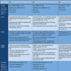
CONCLUSION
A large body of evidence points towards a seminal role for APCs and their uptake receptors in the development of autoimmune diseases. Striking similarities in many genetic and pathophysiological features in autoimmune diseases like SLE, RA and SSc were described despite their apparent dissimilar characteristics (Table 1). A fitting explanation for this issue appears to be the HLA-DRB1 gene which is associated with all these diseases. Since there is almost no overlap between the polymorphisms within this gene [161] and most are located within the epitope binding region, the MHC-II variant probably determines the antigen against which auto-antibodies are produced and thus which clinical phenotype occurs. FcγRs and TLRs expressed by APCs are crucial receptors of the immune system forming a bridge between innate and adaptive immunity. In autoimmunity these receptors appear to direct immune responses towards self by enabling the uptake of immune complexes containing (nucleic) self-antigens. These ICs induce the release of pro-inflammatory cytokines, such as type I IFNs, by APCs and forces them to mature. These mature APCs then induce the differentiation and proliferation of auto-reactive T cells, a crucial event in the pathogenesis of autoimmunity. Next to this the type I IFN system is unequivocally entangled in the pathogenesis of SLE and appears to have contributing roles to both RA and SSc. These findings underscore the potential of treatments aimed at APCs. Accordingly, in the past, present and future the most promising targets in the enduring battle against autoimmunity appear to be these cells themselves, their receptors and their products.