- Home
- About Journals
-
Information for Authors/ReviewersEditorial Policies
Publication Fee
Publication Cycle - Process Flowchart
Online Manuscript Submission and Tracking System
Publishing Ethics and Rectitude
Authorship
Author Benefits
Reviewer Guidelines
Guest Editor Guidelines
Peer Review Workflow
Quick Track Option
Copyediting Services
Bentham Open Membership
Bentham Open Advisory Board
Archiving Policies
Fabricating and Stating False Information
Post Publication Discussions and Corrections
Editorial Management
Advertise With Us
Funding Agencies
Rate List
Kudos
General FAQs
Special Fee Waivers and Discounts
- Contact
- Help
- About Us
- Search


The Open Virology Journal
(Discontinued)
ISSN: 1874-3579 ― Volume 15, 2021
Distribution of Human Papillomavirus Genotypes in Sardinian Patients with Oral Squamous Cell Carcinoma
Caterina Montaldo*, Andrea Mastinu, Stefania Zorco, Noemi Santini, Elisabetta Pisano, Vincenzo Piras, Gloria Denotti, Carla Peluffo, Matteo Erriu, Valentino Garau, Germano Orrù
Abstract
Human papillomaviruses (HPVs) seem to play an important role in the pathogenesis of gynecological carcinomas and in head and neck carcinomas. The aim of this study was to detect and genotype HPVs in fresh oral squamous cell carcinoma (OSCC) from a Sardinian population, and to determine whether HPV presence was significantly associated with the development of OSCC.
The oral mucosa tissues were obtained from 120 samples (68 OSCC and 52 control samples) taken from a Sardinian population seen at the Dental Clinic of the Department of Surgery and Odontostomatological Sciences, University of Cagliari (Italy) and the “ Ospedale SS Trinità”, Cagliari (A.S.L. 8) between 2007 and 2008. PCR was used for the detection of HPV DNA and the genotype was determined by DNA sequencing. The frequency of HPV infection was evaluated in relation to age, sex, smoking and alcohol use. Statistical analysis was performed using the SPSS 11.5 software.
The results showed the presence of HPV-DNA in 60.3% of OSCC with HPV-16 (51.2%) being the most frequent genotype. In these Sardinian OSCC patients, HPV-DNA was detected more in males (65.8%) than in females (34.1%) while controls show a 0% of HPV presence. HPV positive was highly associated with OSCC among subjects with a history of heavy tobacco and alcohol use and among those with no such history.
A greater frequency of high risk HPV presence was observed in patients with OSCC compared to health control patients. In addition these results suggested that oral HPV presence could be associated in OSCC subjects. Our results need more analyses to detect the HPV-DNA integration into tumoral cells.
Article Information
Identifiers and Pagination:
Year: 2010Volume: 4
First Page: 163
Last Page: 168
Publisher Id: TOVJ-4-163
DOI: 10.2174/1874357901004010163
Article History:
Received Date: 1/3/2010Revision Received Date: 15/4/2010
Acceptance Date: 10/5/2010
Electronic publication date: 13/7/2010
Collection year: 2010
open-access license: This is an open access article licensed under the terms of the Creative Commons Attribution Non-Commercial License (http://creativecommons.org/licenses/by-nc/3.0/) which permits unrestricted, non-commercial use, distribution and reproduction in any medium, provided the work is properly cited.
* Address correspondence to this author at the Surgery Department of Odontostomatological Sciences, Odontostomatology Section, O.B.L., University of Cagliari, Via Binaghi n°4, 09121 Cagliari, Italy; Tel: 0039 070.537417; Fax: 0039 070.537437; E-mail: montaldc@unica.it
| Open Peer Review Details | |||
|---|---|---|---|
| Manuscript submitted on 1-3-2010 |
Original Manuscript | Distribution of Human Papillomavirus Genotypes in Sardinian Patients with Oral Squamous Cell Carcinoma | |
INTRODUCTION
Squamous cell carcinoma (SCC) is the most common malignant tumor of the oral cavity and one of the ten most common causes of death. In addition the incidence of oral squamous cell carcinoma (OSCC) of the oral cavity and oropharynx is increasing worldwide [1Nevile BW, Damm DD, Allen CM, Bouqout JE. Oral & Maxillofacial Pathology. 2nd. Philadelphia: WB Sauders 2002; pp. 356-66.]. The annual incidence and mortality rates vary considerably between different races, genders, and age groups; however, as reported in previous publications, the risk of intra-oral cancer rises with increasing age especially for males [1Nevile BW, Damm DD, Allen CM, Bouqout JE. Oral & Maxillofacial Pathology. 2nd. Philadelphia: WB Sauders 2002; pp. 356-66.-4Lawoyin JO, Lawoyin Do, Fasola Ao, Kolude B. Intra-oral squamous cell carcinoma in Nigerians under 40 years of age: a clinicopathological review of eight cases Afr J Med Sci 2005; 34: 99-102.]. Many risk factors have been reported by different authors such as: tobacco use and alcohol consumption which are the main risk factors for this group of cancer [5World cancer report (2003) Lyon, France: International Agency for Research on Cancer. 2003.]. However, these cancers also occur among lifelong tobacco and alcohol abstainers and, thus, other risk factors such as conditions related to oral health and hygiene (i.e. poor condition of the mouth, dentition, bleeding gums, and mouthwash use) have been suspected as contributing to its etiology.
In this context, Human papillomavirus (HPV) as a prognostic risk for OSSC has not yet been extensively studied, a reason for this “defect” could be due to: (i) unusual or less sensitive technologies for HPV detection in SSC, (ii) in excess of 100 HPV genotypes are recognized based on specific DNA sequence variations and 30-40 are associated with sexual lesions. However, the oncogenic capacity of individual HPV types differs considerably [6Gillison ML, Koch WM, Capone RB, et al. Evidence for a causal association between human papillomavirus and a subset of head and neck cancers J Natl Cancer Inst 2000; 92: 709-20.-8Munger K, Howley PM. Human papillomavirus immortalization and transformation functions Virus Res 2002; 89: 213-8.]. This virus is epitheliotropic and in humans is associated with papillomatosis, hyperplastic, and verrucous lesions on the skin and mucous membranes of various sites [9zur Hausen H. Papillomavirus infections—a major cause of human cancers Biochim Biophys Acta 1996; 1288: F55-78.]. These genotypes, which are frequently identified in cellular neoplasm, have been segregated into those with a low risk and those with a high risk of malignant transformation [10Campisi G, Panzarella V, Giuliani M, et al. Human papillomavirus: its identity and controversial role in oral oncogenesis, premalignant and malignant lesions Int J Oncol 2007; 30: 813-23.]. Indeed, some authors have shown that high-risk viral genotypes such as HPV 16, 18, 31 and 33, are frequently associated with leukoplakia and squamous carcinoma [11Ha PK, Califano JA. The role of human papillomavirus in oral carcinogenesis Crit Rev Oral Biol Med 2004; 115: 188-96., 12Furrer VE, Benitez MB, Furnes M, Lanfranchi HE, Modesti NM. Biopsy vs superficial scraping: detection of human papillomavirus 6, 11, 16, and 18 in potentially malignant and malignant oral lesions J Oral Pathol Med 2006; 35: 338-44.]. On the contrary low-risk types, such as HPV 6, 11, 13, 32 are preferentially associated with benign proliferative epithelial lesions e.g. squamous papilloma, condyloma acuminatum, verruca vulgaris and focal epithelial hyperplasia [13Castro TP, Bussoloti Filho I. Prevalence of human papillomavirus (HPV) in oral cavity and oropharynx Braz J Otorhinolaryngol 2006; 72: 272-82.]. Functionally high-risk HPV infection contributes to carcinogenesis and tumor progression through two viral oncogenes E6 and E7 [9zur Hausen H. Papillomavirus infections—a major cause of human cancers Biochim Biophys Acta 1996; 1288: F55-78.]. These oncogenes inhibit the activities of the p53 and pRb, and, have been considered as an important feature in disrupting cell-cycle regulatory pathways leading to a genetic progression to oral squamous cell carcinoma (OSCC) [14zur Hausen H. Papillomaviruses causing cancer: evasion from host-cell control in early events in carcinogenesis J Natl Cancer Inst 2000; 92: 690-8.].
The frequency of HPV in oral lesions varies with geographic occurrence [15Kreimer AR, Clifford GM, Boyle P, Franceschi S. Human papillomavirus types in head and neck squamous cell carcinomas worldwide: a systematic review Cancer Epidemiol Biomarkers Prev 2005; 2: 467-75.], the type of lesion [16Sugiyama M, Bhawal UK, Dohmen T, Ono S, Miyauchi M, Ishikawa T. Detection of human papillomavirus-16 and HPV-18 DNA in normal, dysplastic, and malignant oral epithelium Oral Surg Oral Med Oral Pathol Oral Radiol Endod 2003; 95: 594-600.] and the diagnostic methodology [17Hubbard RA. Human papillomavirus testing methods Arch Pathol Lab Med 2003; 127: 940-5.]. The diagnosis of HPV infection is based on the use of advanced molecular tools [18Husnjak K, Grce M, Magdić L, Pavelić K. Comparison of five different polymerase chain reaction methods for detection of human papillomavirus in cervical cell specimens J Virol Methods 2000; 88: 125-34.]. Polymerase chain reaction (PCR) and sequencing are considered the most sensitive and rapid methods for the detection and genotyping of HPV-DNA, respectively [19Kleter B, van Doorn LJ, Schrauwen L, et al. Development and clinical evaluation of a highly sensitive PCR-reverse hybridization line probe assay for detection and identification of anogenital human papillomavirus J Clin Microbiol 1999; 37: 2508-17.-21Montaldo C, Mastinu A, Quartuccio M, et al. Detection and genotyping of human papillomavirus DNA in samples from healthy Sardinian patients: a preliminary study J Oral Pathol Med 2007; 36: 482-7.].
The present study was conducted to investigate the distribution of HPV in Sardinian patients with malignant oral mucosal lesions compared with healthy patients. An association between HPV-16, HPV -31 exposure and OSCC was also evaluated among subjects with different use of tobacco and alcohol.
MATERIALS AND METHODOLOGY
Clinical Samples
The oral mucosa fresh tissues were obtained from 120 samples (mean age 61.4, age range: 22-86, 69 men and 51 women). Sixty-eight were from the patients with OSCC seen at the “Ospedale SS Trinità”, Cagliari (A.S.L. 8) between 2007 and 2008. Fifty-two control samples were from volunteers randomly chosen from patients at the Department of Dental Disease Prevention (University of Cagliari). These controls reported no history of cancer or showed no sign of mucosal lesions or relevant systemic disease. At the time of examination, all participants were informed about the research and its purpose and gave their informed consent.
An incisional biopsy of the oral squamous cell carcinoma (OSCC) lesion was performed on each patient. All biopsies were divided into two parts: one part was used for histopathological analysis and the other was frozen at -80°C until a molecular assay could be performed. Each fresh specimen sample was analyzed and histopathologically classified as regards to OSCC. Information on patient gender, age, smoking and alcohol history was collected from direct patient interviews and medical records. Tumor grade and staging system (TNM) were obtained from clinical notes, histopathology and radiology reports. Staging was carried out according to the Fifth Edition of the International Cancer Union guidelines [22Sobin LH, Fleming ID. TNM Classification of Malignant Tumors, fifth edition (1997). Union Internationale Contre le Cancer and the American Joint Committee on Cancer Cancer 1997; 80: 1803-4.].
Extraction, Amplification and Sequencing
About 25 mg of tissue was finely crushed and the DNA was extracted with a QIAamp® DNA Mini Kit (Qiagen) according to the manufacturer’s instructions, quantified by spectrophotometry and frozen at -80°C until use. The molecular method for the detection of HPV-DNA was Seminested PCR. In the first reaction the degenerate primers MY09 (5’-CGT CC(AC) AA(AG) GGA (AT)AC TGA TC-3’) and MY11 (5’-GC(AC) CAG GG(AT) CAT AA(CT) AAT GG-3’) were used to amplify a 450 bp fragment in the L1 gene [23Qu W, Jiang G, Cruz Y, et al. PCR detection of human papillomavirus: comparison between MY09/MY11 and GP5+/GP6+ primer systems J Clin Microbiol 1997; 35: 1304-0.]. The first reaction was performed in a final volume of 25 ul, consisting of 4 ul extracted DNA as a template (100 ng), 2.5 ul of a 10X PCR buffer (200 mM Tris-HCl, 500 mM KCl, pH 8.4), 1.5 mM magnesium chloride, a deoxyribonucleotide triphosphate mixture (0.2 mM of each), 0.5 uM of each primers, 2.5 ul U Platinum Taq DNA Polymerase and 4 ul of the Rnasi Dnasi-free template. All reagents were purchased from Invitrogen (Milan, Italy) with the exception of the primers. The mixture was incubated for 2 minutes at 95°C and subjected to 20 cycles of amplification (95°C for 30 seconds, 52°C (MY09/MY11) for 1 minute; and 72°C for 30 seconds) with a final extension at 72°C for 5 minutes in a thermal cycler (Eppendorf, Milan, Italy). Two micro-liters of the PCR product of the first reaction were taken for the PCR second round, with the primer MY09 in conjunction with a new second primer, GP5+N (5’-TTTGTTACTGTGGTAGATAC-3’) to reamplify the material generated in the first round, thus generating a 407 base pair fragment. The second reaction was performed in a final volume of 25 ul and contained 2.5 ul of a 10X PCR buffer (200 mM Tris-HCl, 500 mM KCl, pH 8.4), 1.0 mM magnesium chloride, a deoxyribonucleotide triphosphate mixture (0.2 mM of each), 0.5 uM of each primers and 2.5 ul U Platinum Taq DNA Polymerase. Initial denaturation for 2 minutes at 95°C was followed by 40 cycles. Each of the cycles consisted of denaturation for 30 seconds at 94°C, annealing for 30 seconds at 47°C and chain elongation for 1 minute at 72°C. One cycle of chain elongation was took for 7 minutes at 72°C at the end. The PCR products were electrophoresed on 2% agarose gels, stained with sybr green 1.5% and visualized under an ultraviolet transilluminator. The beta-globin gene was detected (data not shown) in all specimens to confirm the presence of amplifiable DNA in the extracted specimens.
PCR products were purified with a QIAquick Purification PCR kit (Qiagen, Milan, Italy) and were sequenced by a conventional capillary ABI Prism 310 automatic sequencer (Applied Biosystems Foster City, Calif.). The results were edited and analyzed with a Basic Local Alignment Search Tool [24Ladunga I. Finding homologs to nucleotide sequences using network BLAST searches Curr Protoc Bioinformatics 2002. Chap 3: Unit 3.3. Review].
Statistical analysis was performed using the SPSS 11.5 software. Data are expressed as the number of percentages detected. All data were compared using the Chi-Square and Fisher exact with significance considered to be P < 0.05.
RESULTS
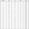
HPV-DNA was detected in 120 tissue samples: 68 oral squamous cell carcinoma and 52 control samples. The age of the patients with OSCC ranged from 27-86 (mean age: 65.6), with 47 males aged between 35-86 (mean age: 61.4) and 21 females aged between 27-84 (mean age: 64.9). The age range of the control patients was 22-85 (mean age: 56.8); 25 males were aged between 40-85 (mean age 57.5) and 27 females between 22-84 (mean age 56.3). Table 1 summarizes the information on HPV-positive patient gender, age. Using the Blast program, the HPV sequences obtained from positive samples showing a representative nucleotide motif were submitted to the DNA data Bank GenBank [24Ladunga I. Finding homologs to nucleotide sequences using network BLAST searches Curr Protoc Bioinformatics 2002. Chap 3: Unit 3.3. Review] with the following accession numbers: DQ286926, DQ312262, DQ312263.
Of the 68 OSCC samples 41 (41/68, 60.3%) were positive for HPV-DNA and indicated by a 407-bp fragment on 2% agarose gel electrophoresis of the PCR products (Fig. 1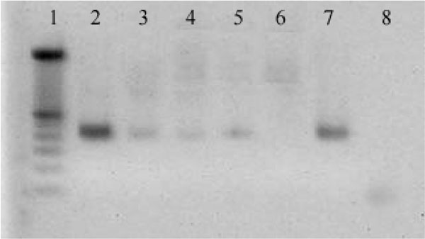 ). Presence of faint bands was read as partial positives that need a confirmation by the sequencing.
 |
DNA from oral tissue was amplified by seminested polymerase chain reaction. Fig. (1). This figure shows the amplicon of the second round reaction generating a 407 bp fragment on 2% agarose gel electrophoresis. Lane 1 is Marker 100 bp, lanes 2, 3, 4 and 5 are positive samples confirmed by nucleotide DNA sequencing, lane 6 is a negative sample. Lane 7 is the positive HPV control and lane 8 is the negative control. Some sequences of HPV types 6 and 31 were submitted to the DNA data Bank (GenBank http://www.ncbi.nlm.nih.gov/BLAST/) with the following accession numbers: DQ286926, DQ312262, DQ312263. The betaglobin gene was detected (data not shown) in all specimens to confirm the presence of amplificable DNA in the extracted specimens. |
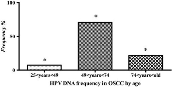 |
Fig. (4). Distribution of HPV in relationship to age, expressed as a frequency percentage. Over 50 years of age a significant increase HPV DNA of the OSCC samples was found. |
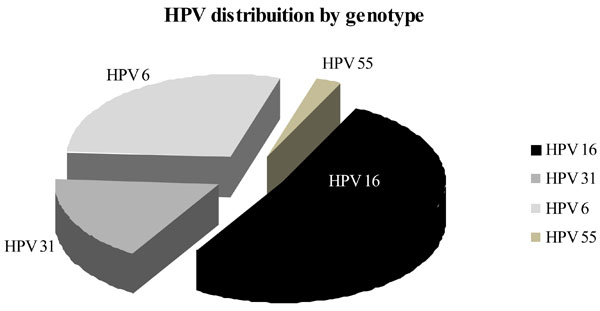
Whereas twenty-eight carcinoma samples (28/41, 68.3%) were positive for a high-risk subtype (21/41, 51.2% HPV-16 and 7/41, 17% HPV-31) only 13 samples out of 41 (34.14%) were positive for low-risk subtype (12/41, 29.3% HPV-6 and 1/41, 2.4% HPV-55) (Fig. 2 ). We were unable to find HPV presence in the 52 samples forming the control group. Moreover, post hoc analysis showed a significant effect for the differences between the OSCC patients and the control group (P < 0.05) for the prevalence of HPV-DNA.
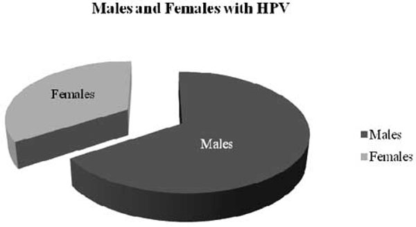
As regards virus distribution between genders (Fig. 3 ), we observed significant differences in HPV frequency between males and females with OSCC from a Sardinian population, 65.8% vs 34.1%, respectively. Furthermore, in subjects over 50 years, a significant increase in HPV DNA (P < 0.05) was detected with advancing age (8% <49 years and 92% >49 years) (Fig. 4
 ).
).
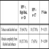
Table 2 shows the lack of statistical associations between the presence of the virus and other known risk factors such as smoking and alcohol. We did not observe a significant difference between HPV high risk presence and tobacco and alcohol use in patients with OSCC.
DISCUSSION
This study was set up to establish the prevalence and HPV genotypes in Sardinian patients with OSCC compared with a group of healthy patients. According to scientific literature, several types of HPV, particularly type 16, have been found to be associated with oropharyngeal squamous-cell carcinoma, a form of head and neck cancer.
The role of HPV in the development of uterine cervix lesions has been demonstrated by many studies [25Tong SY, Lee YS, Park JS, Namkoong SE. Human papillomavirus genotype as a prognostic factor in carcinoma of the uterine cervix Int J Gynecol Cancer 2007; 17: 1307-.]; it still remains a controversial issue as regards oral lesions [26Herrero R, Castellsagué X, Pawlita M, et al. Human papillomavirus and oral cancer: the International Agency for Research on Cancer multicenter study J Natl Cancer Inst 2003; 95: 1772-83.-28Campisi G, Panzarella V, Giuliani M, et al. Human papillomavirus: its identity and controversial role in oral oncogenesis, premalignant and malignant lesions Int J Oncol 2007; 30: 813-23.]. Contradictory results reported on HPV infection in oral mucosa are mainly due to differences in detection methods and the epidemiological characteristics of the observed populations [29Nielsen A, Kjaer SK, Munk C, Iftner T. Type-specific HPV infection and multiple HPV types: prevalence and risk factor profile in nearly 12,000 younger and older Danish women Sex Transm Dis 2008; 35: 276-82., 30Parkin DM, Louie KS, Clifford G. Burden and trends of type-specific human papillomavirus infections and related diseases in the Asia Pacific region Vaccine 2008; 19(26): M1-16.]. Several reports have indeed utilized different molecular techniques (in situ hybridization [31Hikino T. In situ hybridization with novel biotinyl-tyramide: fundamental studies and its utility of the detection of human papilloma virus in tissue sections Rinsho Byori 2007; 55: 922-.] and Southern Blot analysis [32Morgan IM, Taylor ER. Detection and quantitation of HPV DNA replication by Southern blotting and real-time PCR Methods Mol Med 2005; 119: 349-62., 33Mc Kaig RG, Baric RS, Olshan AF. Human papillomavirus and head and neck cancer: epidemiology and molecular biology Head Neck 1998; 20: 250-65.] from several sample typologies (mouthwashes, in scrapings and in paraffin-embedded tissue) [12Furrer VE, Benitez MB, Furnes M, Lanfranchi HE, Modesti NM. Biopsy vs superficial scraping: detection of human papillomavirus 6, 11, 16, and 18 in potentially malignant and malignant oral lesions J Oral Pathol Med 2006; 35: 338-44., 34Lawton G, Thomas S, Schonrock J, Monsour F, Frazer I. Human papillomaviruses in normal oral mucosa: a comparison of methods for sample collection J Oral Pathol Med 1992; 21: 265-9.].
Over the past 20 years, there has been an increasing interest in HPVs as regards their potential role in the pathogenesis of OSCC [35Syrjänen KJ, Syrjänen SM, Lamberg MA, Pyrhönen S. Human papillomavirus (HPV) involvement in squamous cell lesions of the oral cavity Proc Finn Dent Soc 1983; 79: 1-8.-41Trottier H, Franco EL. Human papillomavirus and cervical cancer: burden of illness and basis for prevention Am J Manag Care 2006; 12: 462-72.]. Nevertheless, a recent paper has confirmed HPV as an independent risk factor for OSCC compared to other known factors such as alcohol and smoking [42Miller CS, White DK. Human papillomavirus expression in oral mucosa, premalignant conditions, and squamous cell carcinoma: a retrospective review of the literature Oral Surg Oral Med Oral Pathol Oral Radiol Endod 1996; 82: 57-68.]. Several HPV genotypes (1, 2, 3, 4, 6, 7, 10, 11, 13, 16, 18, 31, 32, 33, 35, 55, 57) have been isolated from oral lesions [12Furrer VE, Benitez MB, Furnes M, Lanfranchi HE, Modesti NM. Biopsy vs superficial scraping: detection of human papillomavirus 6, 11, 16, and 18 in potentially malignant and malignant oral lesions J Oral Pathol Med 2006; 35: 338-44., 43Anaya-Saavedra G, Ramírez-Amador V, Irigoyen-Camacho ME, et al. High association of human papillomavirus infection with oral cancer: a case-control study Arch Med Res 2008; 39: 189-97.-45Schwartz SR, Yueh B, McDougall JK, Daling JR, Schwartz SM. Human papillomavirus infection and survival in oral squamous cell cancer: a population-based study Otolaryngol Head Neck Surg 2001; 125: 1-9.]. However, in OSCC, the most commonly HPV genotype detected is 16, which has been demonstrated in 90-95% of all HPV positive neoplasms, followed by HPV 18 and HPV 31 [45Schwartz SR, Yueh B, McDougall JK, Daling JR, Schwartz SM. Human papillomavirus infection and survival in oral squamous cell cancer: a population-based study Otolaryngol Head Neck Surg 2001; 125: 1-9.-47Smith EM, Ritchie JM, Summersgill KF, et al. Age, sexual behavior and human papillomavirus infection in oral cavity and oropharyngeal cancers Int J Cancer 2004; 108: 766-2.].
This study was performed on a Sardinian population, which presents genetic homogeneity compared to other known populations in Europe, including the Italian population [48Barbujani G, Sokal RR. Genetic population structure of Italy. Geographic patterns of gene frequencies Hum Biol 1991; 63: 253-72.]. Furthermore, a highly sensitive/specific molecular methodology such as nested PCR followed by capillary sequencing was used [49Lin H, Moh JS, Ou YC, et al. A simple method for the detection and genotyping of high-risk human papillomavirus using seminested polymerase chain reaction and reverse hybridization Gynecol Oncol 2005; 96: 84-91.]. These results confirm the prevalence of HPV-DNA in OSCC and also that HPV 16 is the most frequent genotype. The findings of the present study are in agreement with those reported by Miller et al. [50Miller CS, Johnstone BM. Human papillomavirus as a risk factor for oral squamous cell carcinoma: a meta-analysis, 1982-1997 Oral Surg Oral Med Oral Pathol Oral Radiol Endod 2001; 91: 622-35.] and Anaya-Saavedra et al. [43Anaya-Saavedra G, Ramírez-Amador V, Irigoyen-Camacho ME, et al. High association of human papillomavirus infection with oral cancer: a case-control study Arch Med Res 2008; 39: 189-97.] in two recent studies, suggesting that HPV 16 represents a risk factor in the etiopathogenesis of a subset of OSCC.
Despite the presence of low-risk, HPV (29.3% HPV-6 and 2.4% HPV-55) is according to the results of Herrero et al. [26Herrero R, Castellsagué X, Pawlita M, et al. Human papillomavirus and oral cancer: the International Agency for Research on Cancer multicenter study J Natl Cancer Inst 2003; 95: 1772-83.] and other authors [44Chang F, Syrjänen S, Kellokoski J, Syrjänen K. Human papillomavirus (HPV) infections and their associations with oral disease J Oral Pathol Med 1991; 20: 305-17., 51Go C, Schwartz MR, Donovan DT. Molecular transformation of recurrent respiratory papillomatosis: viral typing and p53 overexpression Ann Otol Rhinol Laryngol 2003; 112: 298-302., 52Reidy PM, Dedo HH, Rabah R, et al. Integration of human papillomavirus type 11 in recurrent respiratory papilloma-associated cancer Laryngoscope 2004; 114: 1906-09.] who have demonstrated a malignant degeneration of papillomas induced by low-risk HPV. For example Chang et al. [44Chang F, Syrjänen S, Kellokoski J, Syrjänen K. Human papillomavirus (HPV) infections and their associations with oral disease J Oral Pathol Med 1991; 20: 305-17.] reported that HPV 6 was also found in squamous cell carcinoma. In addition the same bibliographic data showed that an association between multiple infection (HPV 6, HPV 11 and HPV 16) is required for the development of malignant lesions [39Bouda M, Gorgoulis VG, Kastrinakis NG, et al. "High risk" HPV types are frequently detected in potentially malignant and malignant oral lesions, but not in normal oral mucosa Mod Pathol 2000; 13: 644-53.]. Our results give us the basis to further determine how many virus’ presence could be fundamental in the carcinogenesis process through the study of the HPV DNA integration.
In our study we observed a different frequency between genders; in fact a major presence of HPV-DNA results in males compared to females, as has been described in previous studies [53Marais DJ, Sampson C, Jeftha A, et al. More men than women make mucosal IgA antibodies to Human papillomavirus type 16 (HPV-16) and HPV-18: a study of oral HPV and oral HPV antibodies in a normal healthy population BMC Infect Dis 2006; 6: 95.]. Moreover, this finding indicates that higher positivity may be the result of accumulated lifetime exposure to HPVs. The oral cavity is the anatomic site with the most frequent exposure to viruses and we observed an increase in HPV positivity with age (Fig. 4) in accordance to many papers [47Smith EM, Ritchie JM, Summersgill KF, et al. Age, sexual behavior and human papillomavirus infection in oral cavity and oropharyngeal cancers Int J Cancer 2004; 108: 766-2., 54Cruz IB, Snijders PJ, Steenbergen RD, et al. Age-dependence of human papillomavirus DNA presence in oral squamous cell carcinomas Eur J Cancer B Oral Oncol 1996; 1: 55-62., 55Dunne EF UE, Sternberg M, McQuillan G, Swan DC, Patel SS, Markowitz LE. Prevalence of HPV infection among females in the United States JAMA 2007; 28: 809-13.]. This increment may be explained by the fact that different factors can be involved in the malignant process such as: immune defences [56Lesourd B. Nutritional factors and immunological ageing Proc Nutr Soc 2006; 65: 319-25.], nutritional deficiencies [57Bologna-Molina RE, Castaneda-Castaneira RE, Molina-Frechero N, Perez-Rodriguez E. Human papilloma virus and its association with oral cancer Rev Med Inst Mex Seguro Soc 2006; 44: 147-53.], traumatisms inflicted by fractured teeth, incongruous restorations or prostheses [58Perez MA, Raimondi AR, Itoiz ME. An experimental model to demonstrate the carcinogenic action of oral chronic traumatic ulcer J Oral Pathol Med 2005; 34: 17-22., 59Czerninski R, Kaplan I, Almoznino G, Maly A, Regev E. Oral squamous cell carcinoma around dental implants Quintessence Int 2006; 37: 707-11.], and poor oral hygiene [60Rosenquist K. Risk factors in oral and oropharyngeal squamous cell carcinoma: a population-based case-control study in southern Sweden Swed Dent J Suppl 2005; 179: 1-66.] which worsens with an increase in age. Moreover, according to our findings, HPV is a specific risk factor for the development of OSCC in association with other important risks i.e. smoking and alcohol [61Boffetta P, Mashberg A, Winkelmann R. Carcinogenic effect of tobacco smoking and alcohol drinking on anatomic sites of the oral cavity and oropharynx Int J Cancer 1992; 52: 530-3.-65Farshadpour F, Hordijk GJ, Koole R, Slootweg PJ. Non-smoking and non-drinking patients with head and neck squamous cell carcinoma: a distinct population Oral Dis 2007; 13: 239-43.].
CONCLUSION
Our data support the presence of high frequency of HPV 16 in oral cancer, but also suggest the possibility of an involvement of low-risk HPVs as a cofactor in this malignant process; in this context the age of the patients plays a central role in the cancer onset. Moreover, the results of this study, probably because of small sample size, did not show the association significantly among HPV and tobacco and alcohol use. Next step, to verify the real HPV role in carcinogenesis, will be the study of the possible HPV infection determined by the integration in the human genome confirmed by the study of E6 and E7 mRNA expression [14zur Hausen H. Papillomaviruses causing cancer: evasion from host-cell control in early events in carcinogenesis J Natl Cancer Inst 2000; 92: 690-8.].
ACKNOWLEDGEMENTS
This work was funded by Italian MIUR (Ministry of the Instruction, University and Research) by PRIN 2003 Programs of Research National Interest.